
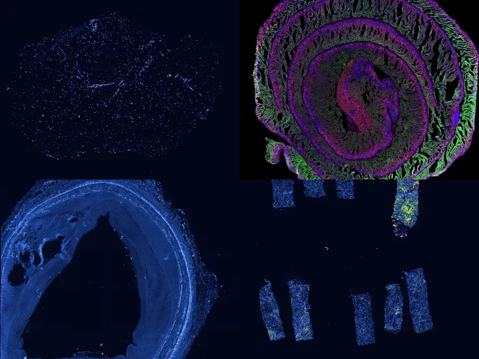
Human kidney
GSM8933095
human kidney 6.3 GB 307,653 cells bronze NCBI GEO policy
Technology10x Xenium
Organismhuman
Tissuekidney
Cells307,653
bronze NCBI GEO policy
Rapidly progressive glomerulonephritis (RPGN) is the most aggressive group of autoimmune kidney disease with the worst prognosis. Anti-neutrophil cytoplasmic antibody (ANCA) associated vasculitis, anti-glomerular basement membrane (anti-GBM) and lupus nephritis are the most common causes of RPGN and are characterized by the formation of glomerular crescents and infiltration of leukocytes that eventually lead to glomerulosclerosis and kidney failure. In this work, we used high-resolution spatial transcriptomics of 32 ANCA, 19 lupus nephritis, 6 anti-GBM, and 6 control patients to understand how intercellular signaling between immune and renal tissue cells leads to renal inflammation and glomerular injury. Using 3,218,210 immune and kidney cells, we observed that the biological pathways involved in the sequence of glomerular crescent formation are similar across the diseases. While innate immune cells infiltrated the glomerular compartment relatively early, later increases in adaptive immune cells were largely restricted to the periglomerular regions. These changes in immune cells temporally correlated with increases in glomerular parietal epithelial (PEC) and fibrotic mesangial cells, suggesting disease-relevant functional signaling between these immune and renal cells. Cell communication analysis revealed early disease PDGF signaling from epithelial and mesangial cells to PECs, causing their activation and proliferation. At later stages, TGF-β signaling from macrophages, T cells, epithelial cells, and mesangial cells to PECs triggered the expression of extracellular matrix components resulting in glomerulosclerosis. Our results highlight a spatio-temporally conserved progression into glomerular crescents and sclerosis for ANCA, lupus nephritis, and anti-GBM disease, which is driven by consecutive PDGF and TGF-β signaling to PECs.
from sddb import Catalog

cohort = Catalog().collection("geo-gse294965")
cohort.members        # the 9 datasets as a table

print(cohort.licenses())              # check the terms before you download

sdatas = cohort.to_dict()             # {uid: SpatialData}, cached locally
# or keep the .zarr files in a folder of your choice:
# sdatas = cohort.to_dict(dest="data/")Needs pip install lamindb and network access to the Lamin database, which is limited during the alpha. If it fails, use the Client tab.
import lamindb as ln

db = ln.DB("scverse/spatialdata-db")
coll = db.Collection.get(key="geo-gse294965")
coll.ordered_artifacts.to_dataframe()   # the 9 members, ordered

# download every member .zarr to the local cache:
paths = [a.cache() for a in coll.ordered_artifacts.all()]# sddb client: download every member .zarr into data/
sddb-cli download --collection geo-gse294965 --dest data/Licenses: 9 NCBI GEO policy
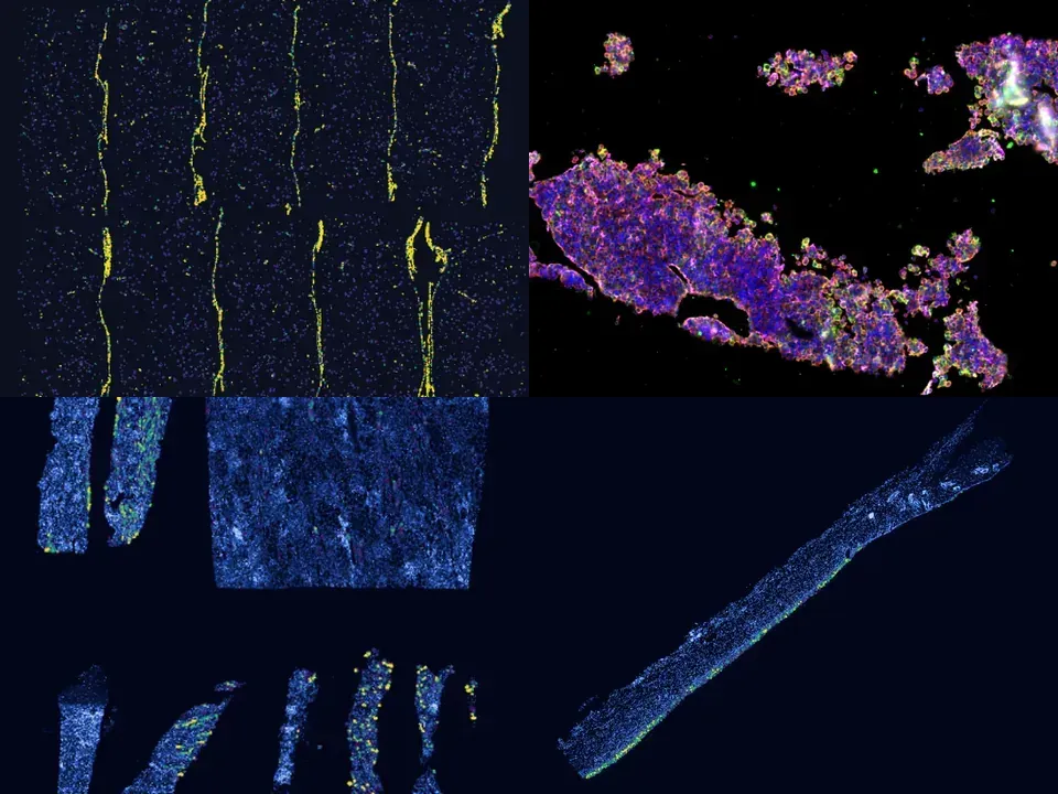
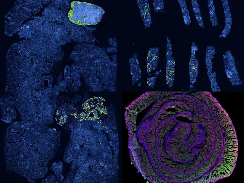
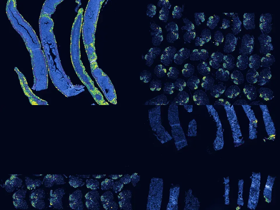
In anti-neutrophil cytoplasmic antibody-associated vasculitis (AAV) and systemic lupus erythematosus (SLE), glomerulonephritis is a severe kidney complication driven by immune cells, including T cells. However, the mechanisms underlying T cell activation in these contexts remain elusive. Here we report that in patients with AAV and SLE, type I interferon (IFN-I) induces T cell differentiation into interferon-stimulated genes-expressing T (ISG-T) cells, which are characterized by an elevated IFN-I signature, an immature phenotype, and cytotoxicity in inflamed tissue. Mechanistically, IFN-I stimulates the expression of interferon regulatory factor 7 (IRF7) in T cells, which in turn induces granzyme B production. In mice, blocking IFN-I signaling reduces IRF7 and granzyme B expression in T cells, thus ameliorating glomerulonephritis. In parallel, spatial transcriptomic analyses of kidney biopsies from patients with AAV or SLE reveal an elevated ISG signature and the presence of ISG-T cells in close proximity to plasmacytoid dendritic cells, the primary producers of IFN-I. Our results from both patients and animal models thus suggest that IFN-I production in inflamed tissue may drive ISG-T cell differentiation to expand the pool of cytotoxic T cells in autoimmune diseases.
@article{Wang_2025,
  title = {Type I interferon drives T cell cytotoxicity by upregulation of interferon regulatory factor 7 in autoimmune kidney diseases in mice},
  volume = {16},
  ISSN = {2041-1723},
  url = {http://dx.doi.org/10.1038/s41467-025-59819-7},
  DOI = {10.1038/s41467-025-59819-7},
  number = {1},
  journal = {Nature Communications},
  publisher = {Springer Science and Business Media LLC},
  author = {Wang, Huiying and Engesser, Jonas and Khatri, Robin and Schaub, Darius P. and Paust, Hans-Joachim and Sultana, Zeba and Jauch-Speer, Saskia-Larissa and Peters, Anett and Kaffke, Anna and Bonn, Stefan and Huber, Tobias B. and Mittrücker, Hans-Willi and Krebs, Christian F. and Panzer, Ulf and Asada, Nariaki},
  year = {2025},
  month = May
}