
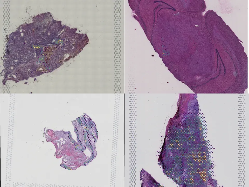
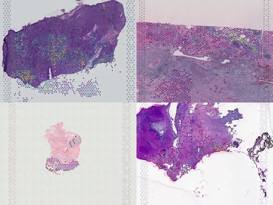
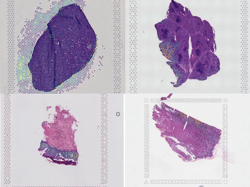
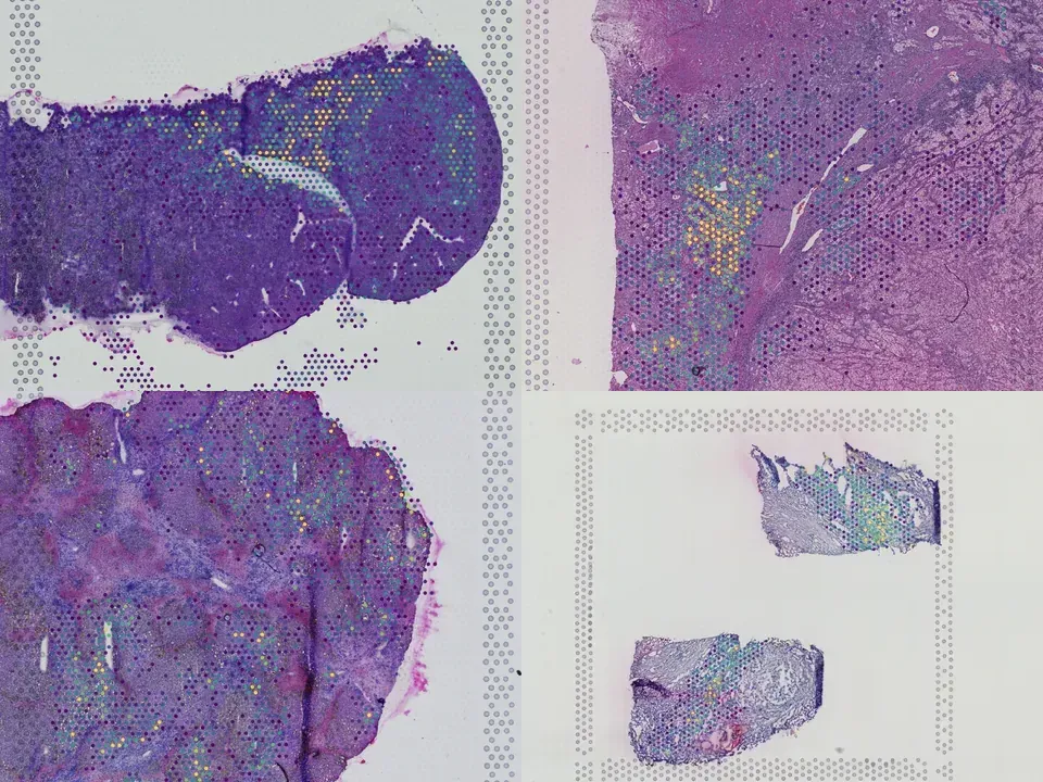
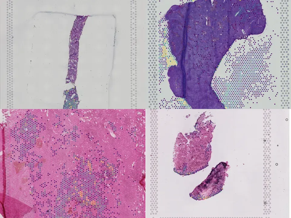
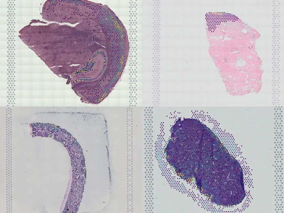
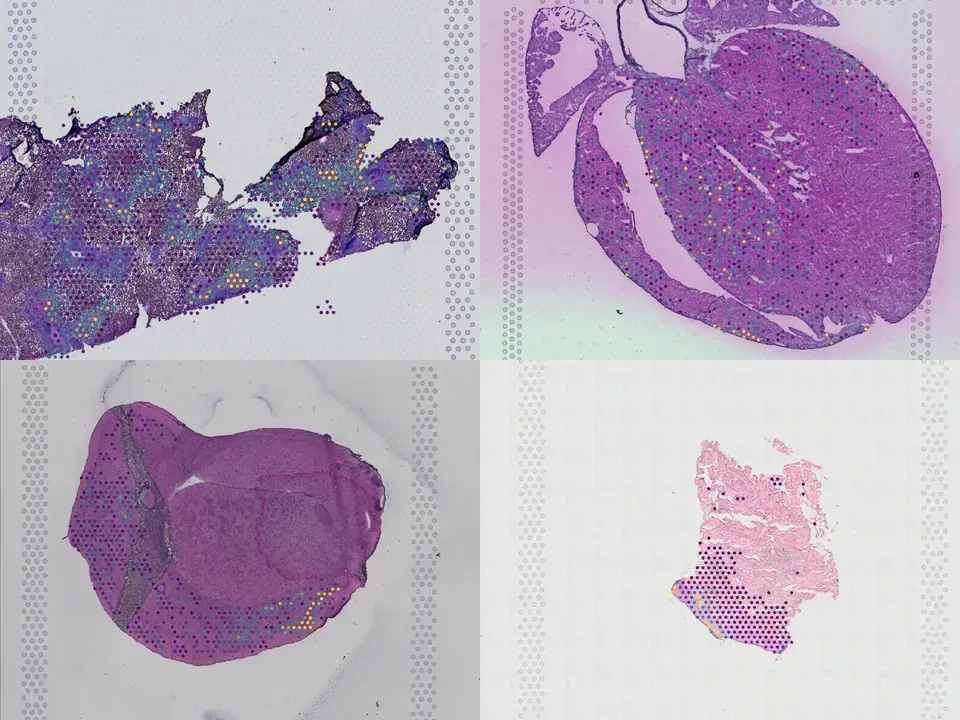
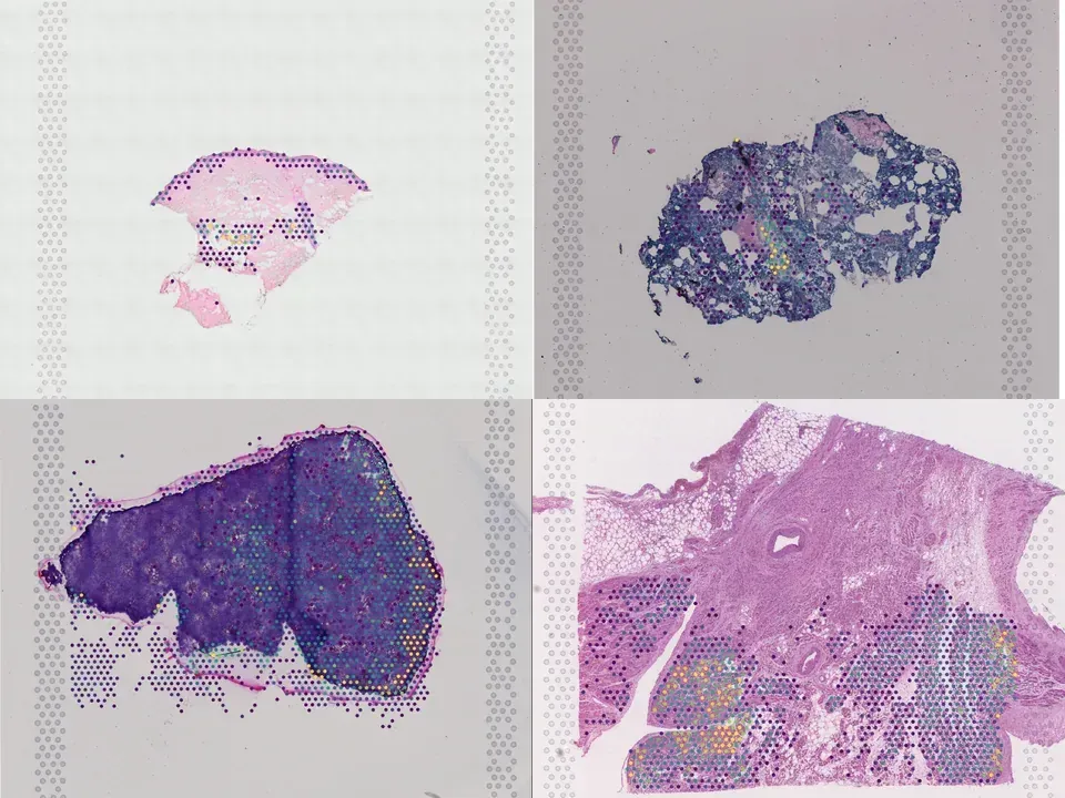
Human liver
NCBI830
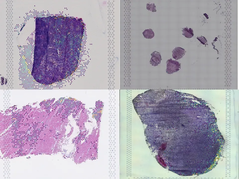
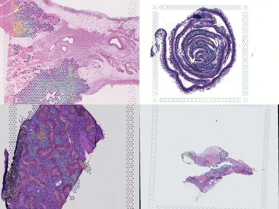
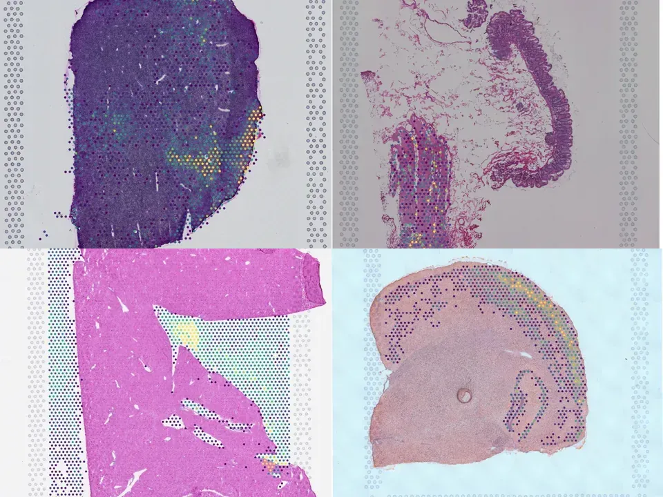
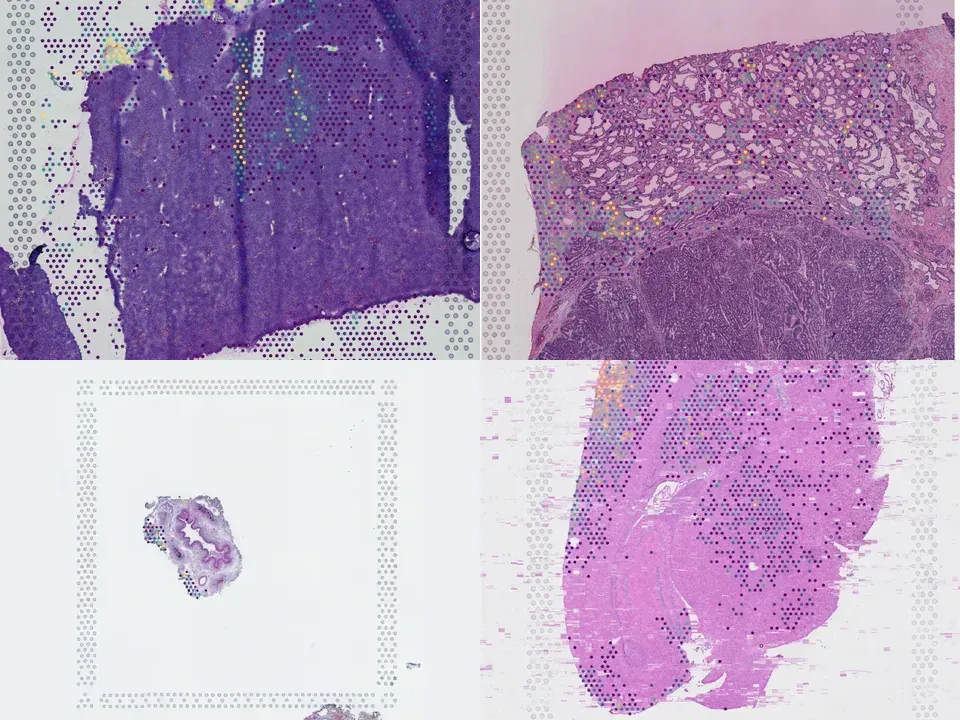
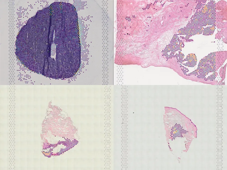
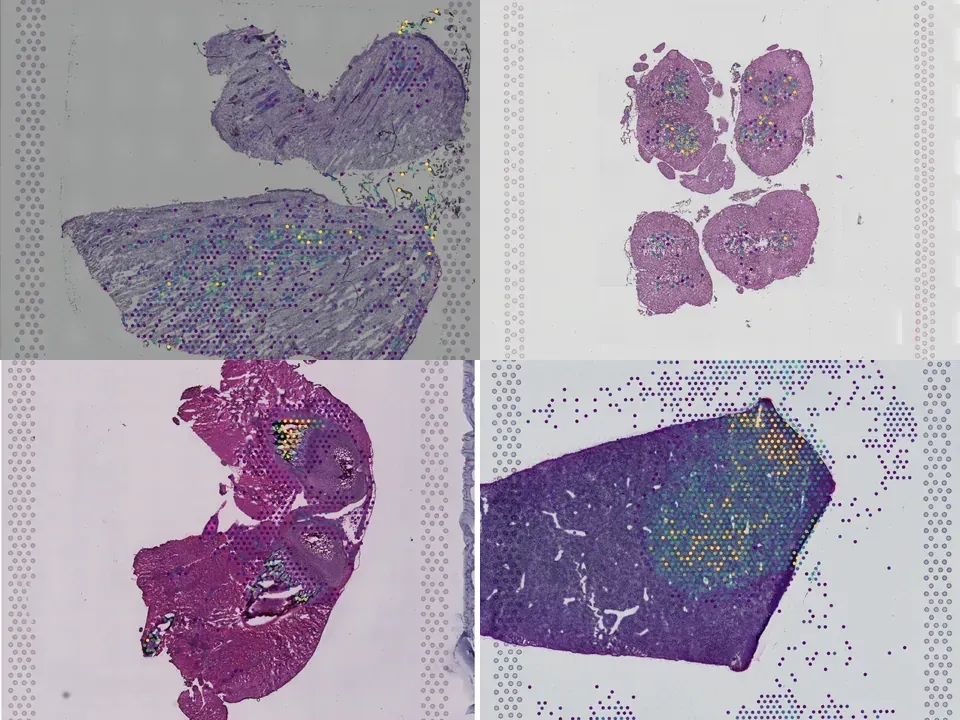
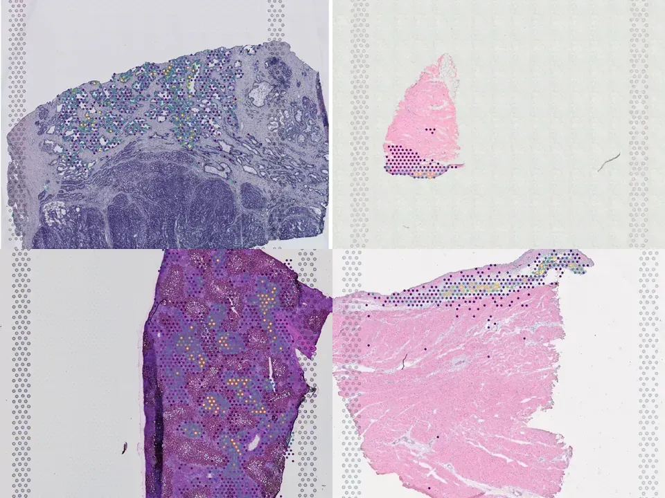
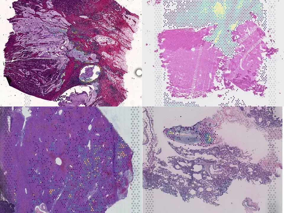
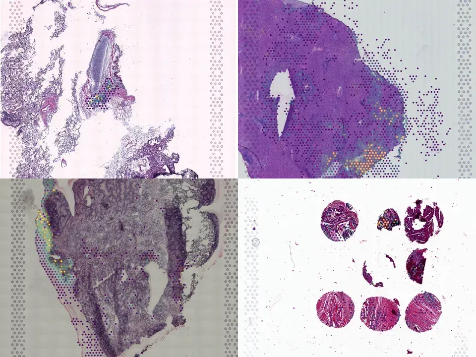
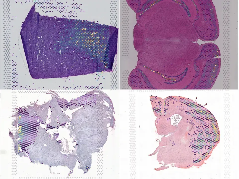
human liver 162 MB 4,992 spots silver H&E
TechnologyVisium
Organismhuman
Tissueliver
Spots4,992
silver H&E
from sddb import Catalog

cohort = Catalog().collection("doi-10-1038-s41586-024-07376-2")
cohort.members        # the 19 datasets as a table

print(cohort.licenses())              # check the terms before you download

sdatas = cohort.to_dict()             # {uid: SpatialData}, cached locally
# or keep the .zarr files in a folder of your choice:
# sdatas = cohort.to_dict(dest="data/")Needs pip install lamindb and network access to the Lamin database, which is limited during the alpha. If it fails, use the Client tab.
import lamindb as ln

db = ln.DB("scverse/spatialdata-db")
coll = db.Collection.get(key="doi-10.1038/s41586-024-07376-2")
coll.ordered_artifacts.to_dataframe()   # the 19 members, ordered

# download every member .zarr to the local cache:
paths = [a.cache() for a in coll.ordered_artifacts.all()]# sddb client: download every member .zarr into data/
sddb-cli download --collection doi-10-1038-s41586-024-07376-2 --dest data/Licenses: 19 CC BY-NC-SA 4.0
The liver has a unique ability to regenerate<sup>1,2</sup>; however, in the setting of acute liver failure (ALF), this regenerative capacity is often overwhelmed, leaving emergency liver transplantation as the only curative option<sup>3-5</sup>. Here, to advance understanding of human liver regeneration, we use paired single-nucleus RNA sequencing combined with spatial profiling of healthy and ALF explant human livers to generate a single-cell, pan-lineage atlas of human liver regeneration. We uncover a novel ANXA2<sup>+</sup> migratory hepatocyte subpopulation, which emerges during human liver regeneration, and a corollary subpopulation in a mouse model of acetaminophen (APAP)-induced liver regeneration. Interrogation of necrotic wound closure and hepatocyte proliferation across multiple timepoints following APAP-induced liver injury in mice demonstrates that wound closure precedes hepatocyte proliferation. Four-dimensional intravital imaging of APAP-induced mouse liver injury identifies motile hepatocytes at the edge of the necrotic area, enabling collective migration of the hepatocyte sheet to effect wound closure. Depletion of hepatocyte ANXA2 reduces hepatocyte growth factor-induced human and mouse hepatocyte migration in vitro, and abrogates necrotic wound closure following APAP-induced mouse liver injury. Together, our work dissects unanticipated aspects of liver regeneration, demonstrating an uncoupling of wound closure and hepatocyte proliferation and uncovering a novel migratory hepatocyte subpopulation that mediates wound closure following liver injury. Therapies designed to promote rapid reconstitution of normal hepatic microarchitecture and reparation of the gut-liver barrier may advance new areas of therapeutic discovery in regenerative medicine.
@article{Matchett_2024,
  title = {Multimodal decoding of human liver regeneration},
  volume = {630},
  ISSN = {1476-4687},
  url = {http://dx.doi.org/10.1038/s41586-024-07376-2},
  DOI = {10.1038/s41586-024-07376-2},
  number = {8015},
  journal = {Nature},
  publisher = {Springer Science and Business Media LLC},
  author = {Matchett, K. P. and Wilson-Kanamori, J. R. and Portman, J. R. and Kapourani, C. A. and Fercoq, F. and May, S. and Zajdel, E. and Beltran, M. and Sutherland, E. F. and Mackey, J. B. G. and Brice, M. and Wilson, G. C. and Wallace, S. J. and Kitto, L. and Younger, N. T. and Dobie, R. and Mole, D. J. and Oniscu, G. C. and Wigmore, S. J. and Ramachandran, P. and Vallejos, C. A. and Carragher, N. O. and Saeidinejad, M. M. and Quaglia, A. and Jalan, R. and Simpson, K. J. and Kendall, T. J. and Rule, J. A. and Lee, W. M. and Hoare, M. and Weston, C. J. and Marioni, J. C. and Teichmann, S. A. and Bird, T. G. and Carlin, L. M. and Henderson, N. C.},
  year = {2024},
  month = May,
  pages = {158–165}
}