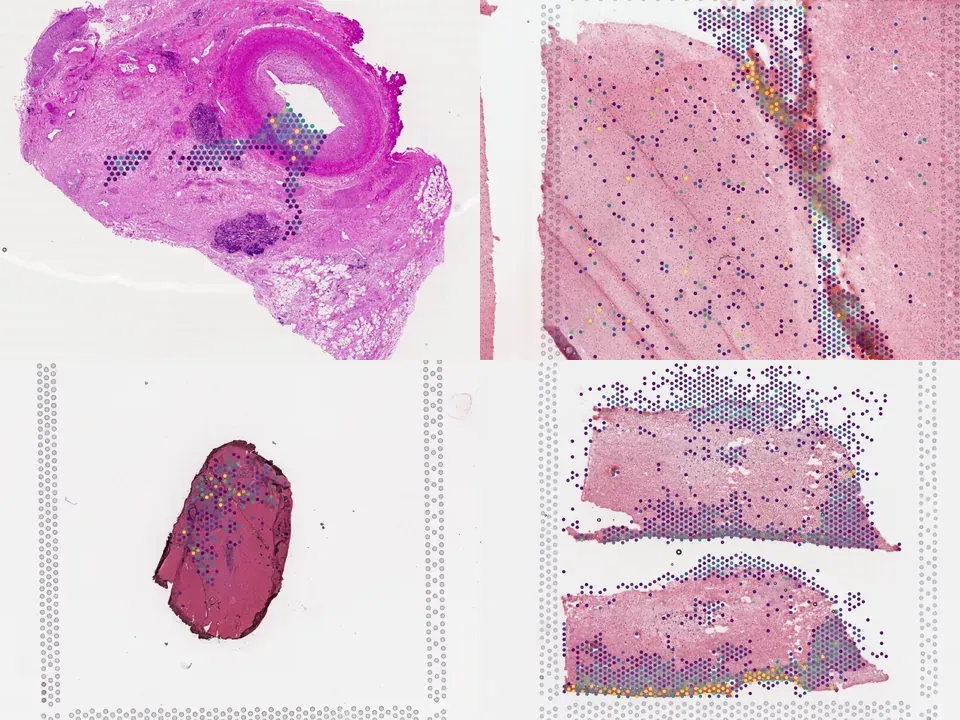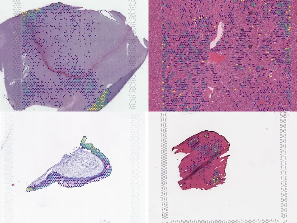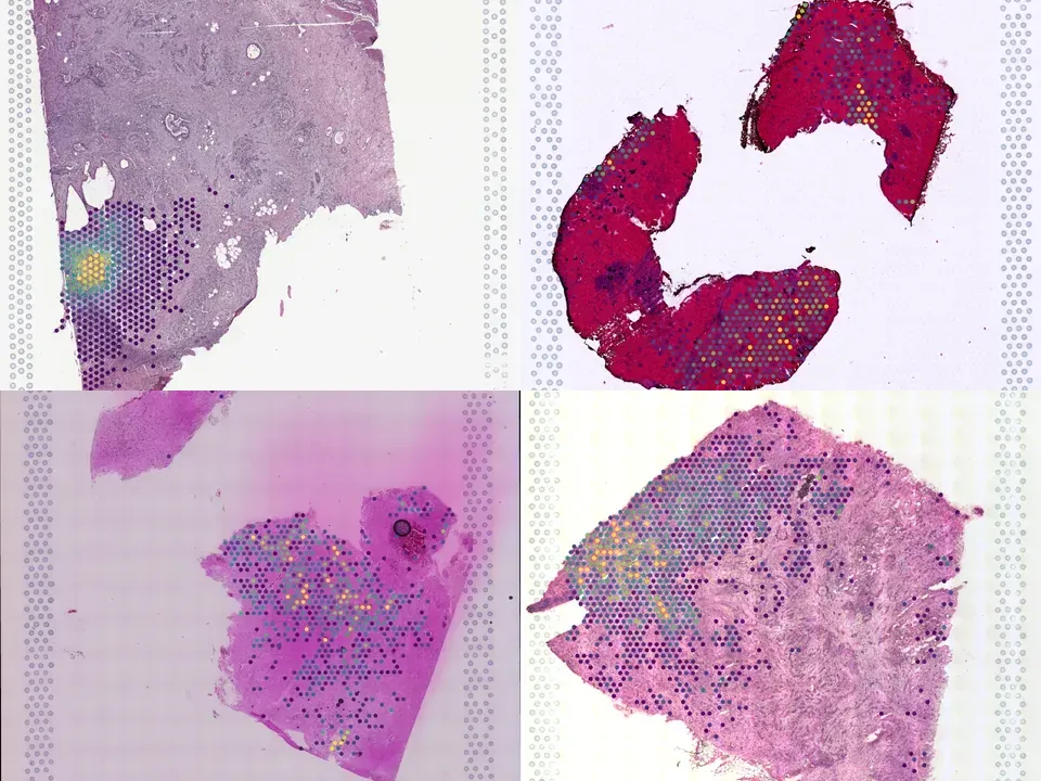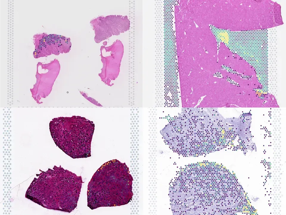
Collections / Spatiotemporal transcriptomic mapping of regenerative inflammation in skeletal muscle reveals a dynamic multilayered tissue architecture

Spatiotemporal transcriptomic mapping of regenerative inflammation in skeletal muscle reveals a dynamic multilayered tissue architecture

Tissue regeneration is orchestrated by macrophages that clear damaged cells and promote regenerative inflammation. How macrophages spatially adapt and diversify their functions to support the architectural requirements of actively regenerating tissue remains unknown. In this study, we reconstructed the dynamic trajectories of myeloid cells isolated from acutely injured and early-stage dystrophic muscles. We identified divergent subsets of monocytes/macrophages and dendritic cells (DCs) and validated markers (e.g., GPNMB) and transcriptional regulators associated with defined functional states. In dystrophic muscle, specialized repair-associated subsets exhibited distinct macrophage diversity and reduced DC heterogeneity. Integrating spatial transcriptomics analyses with immunofluorescence uncovered the ordered distribution of subpopulations and multilayered regenerative inflammation zones (RIZs) where distinct macrophage subsets are organized in functional zones around damaged myofibers supporting all phases of regeneration. Importantly, intermittent glucocorticoid treatment disrupted the RIZs. Our findings suggest that macrophage subtypes mediated the development of the highly ordered architecture of regenerative tissues, unveiling the principles of the structured yet dynamic nature of regenerative inflammation supporting effective tissue repair.

Datasets7
Size~101 MB
AssayVisium
Samplemouse · multiple
KindComparison
Flag a data issue
PMID:39190487 ↗
Python · sddb client
from sddb import Catalog

cohort = Catalog().collection("geo-gse223813")
cohort.members        # the 7 datasets as a table

print(cohort.licenses())              # check the terms before you download

sdatas = cohort.to_dict()             # {uid: SpatialData}, cached locally
# or keep the .zarr files in a folder of your choice:
# sdatas = cohort.to_dict(dest="data/")
Python · lamindb

Needs pip install lamindb and network access to the Lamin database, which is limited during the alpha. If it fails, use the Client tab.

import lamindb as ln

db = ln.DB("scverse/spatialdata-db")
coll = db.Collection.get(key="geo-gse223813")
coll.ordered_artifacts.to_dataframe()   # the 7 members, ordered

# download every member .zarr to the local cache:
paths = [a.cache() for a in coll.ordered_artifacts.all()]
Shell
# sddb client: download every member .zarr into data/
sddb-cli download --collection geo-gse223813 --dest data/

Licenses: 7 NCBI GEO policy

Open in Vitessce
Visium
Mouse gastrocnemius
GSM6996264
mouse gastrocnemius Duchenne muscular dystrophy 15.2 MB 814 spots silver H&E NCBI GEO policy
TechnologyVisium
Organismmouse
Tissuegastrocnemius
ConditionDuchenne muscular dystrophy
Size15.2 MB
Spots814
silver H&E NCBI GEO policy Open in Vitessce
Open in Vitessce
Visium
Mouse gastrocnemius
GSM6996262
mouse gastrocnemius Duchenne muscular dystrophy 9.6 MB 653 spots silver H&E NCBI GEO policy
TechnologyVisium
Organismmouse
Tissuegastrocnemius
ConditionDuchenne muscular dystrophy
Size9.6 MB
Spots653
silver H&E NCBI GEO policy Open in Vitessce
Open in Vitessce
Visium
Mouse gastrocnemius
GSM6996261
mouse gastrocnemius Duchenne muscular dystrophy 12.1 MB 856 spots silver H&E NCBI GEO policy
TechnologyVisium
Organismmouse
Tissuegastrocnemius
ConditionDuchenne muscular dystrophy
Size12.1 MB
Spots856
silver H&E NCBI GEO policy Open in Vitessce
Open in Vitessce
Visium
Mouse gastrocnemius
GSM6996263
mouse gastrocnemius Duchenne muscular dystrophy 19.6 MB 1,589 spots silver H&E NCBI GEO policy
TechnologyVisium
Organismmouse
Tissuegastrocnemius
ConditionDuchenne muscular dystrophy
Size19.6 MB
Spots1,589
silver H&E NCBI GEO policy Open in Vitessce
Visium
Mouse tibialis anterior
GSM6996258
mouse tibialis anterior 9.7 MB 770 spots silver H&E NCBI GEO policy
TechnologyVisium
Organismmouse
Tissuetibialis anterior
Size9.7 MB
Spots770
silver H&E NCBI GEO policy
Open in Vitessce
Visium
Mouse tibialis anterior
GSM6996260
mouse tibialis anterior 18.3 MB 1,567 spots silver H&E NCBI GEO policy
TechnologyVisium
Organismmouse
Tissuetibialis anterior
Size18.3 MB
Spots1,567
silver H&E NCBI GEO policy Open in Vitessce
Open in Vitessce
Visium
Mouse tibialis anterior
GSM6996259
mouse tibialis anterior 16.8 MB 1,273 spots silver H&E NCBI GEO policy
TechnologyVisium
Organismmouse
Tissuetibialis anterior
Size16.8 MB
Spots1,273
silver H&E NCBI GEO policy Open in Vitessce

Cite this collection

No linked publication found — please cite the data source.