
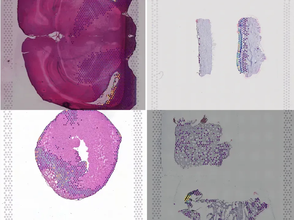
Human eye
NCBI592
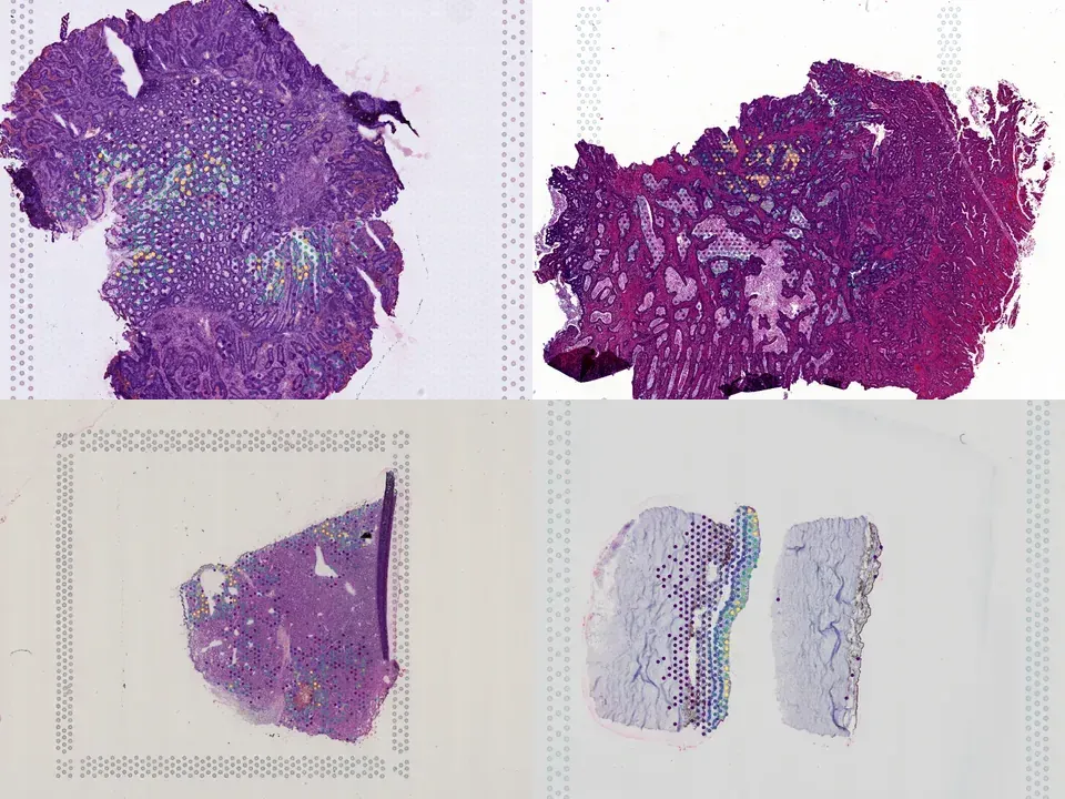
human eye wet macular degeneration 434 MB 721 spots bronze H&E
TechnologyVisium
Organismhuman
Tissueeye
Conditionwet macular degeneration
Spots721
Magnification20x · 0.465 µm/px
bronze H&E
from sddb import Catalog

cohort = Catalog().collection("doi-10-1101-2023-06-16-544770")
cohort.members        # the 5 datasets as a table

print(cohort.licenses())              # check the terms before you download

sdatas = cohort.to_dict()             # {uid: SpatialData}, cached locally
# or keep the .zarr files in a folder of your choice:
# sdatas = cohort.to_dict(dest="data/")Needs pip install lamindb and network access to the Lamin database, which is limited during the alpha. If it fails, use the Client tab.
import lamindb as ln

db = ln.DB("scverse/spatialdata-db")
coll = db.Collection.get(key="doi-10.1101/2023.06.16.544770")
coll.ordered_artifacts.to_dataframe()   # the 5 members, ordered

# download every member .zarr to the local cache:
paths = [a.cache() for a in coll.ordered_artifacts.all()]# sddb client: download every member .zarr into data/
sddb-cli download --collection doi-10-1101-2023-06-16-544770 --dest data/Licenses: 5 CC BY-NC-SA 4.0
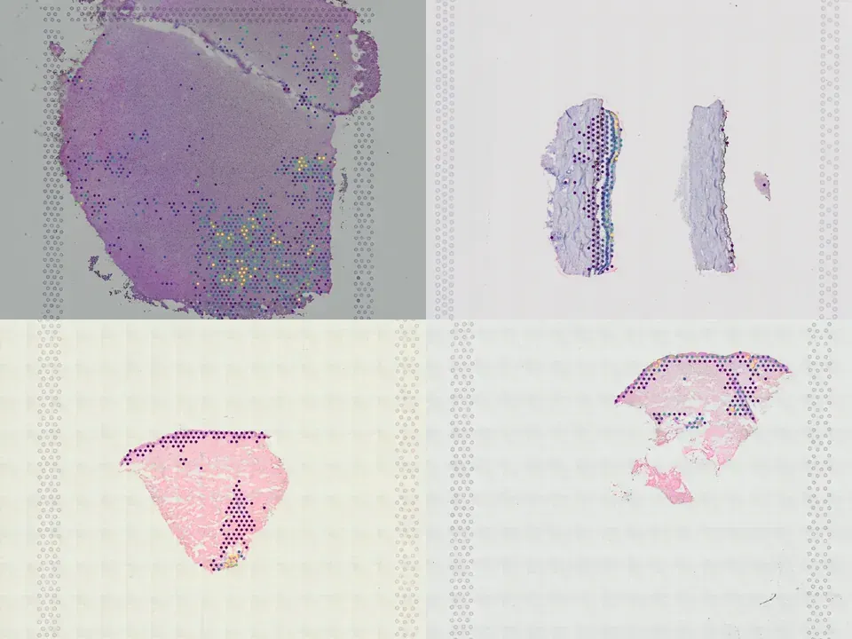
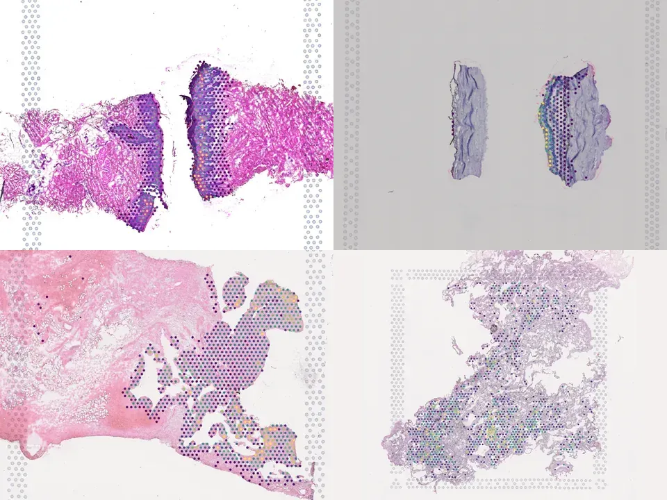
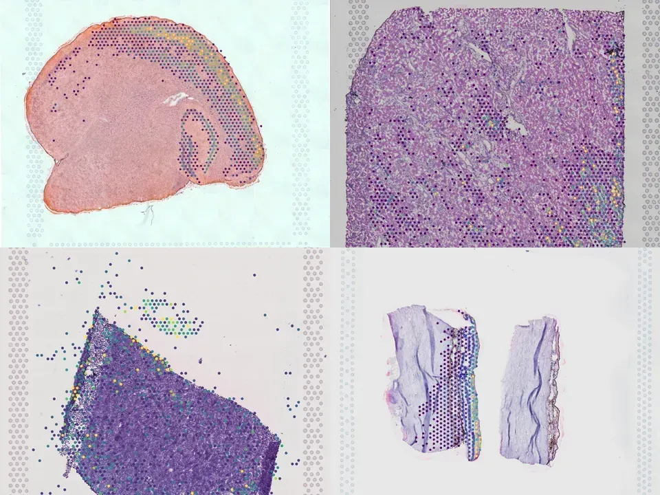
<h4>ABSTRACT</h4> Macular neovascularization is a relatively common and potentially visually devastating complication of age-related macular degeneration. In macular neovascularization, pathologic angiogenesis can originate from either the choroid or the retina, but we have limited understanding of how different cell types become dysregulated in this dynamic process. In this study, we performed spatial RNA sequencing on a human donor eye with macular neovascularization as well as a healthy control donor. We identified genes enriched within the area of macular neovascularization and used deconvolution algorithms to predict the originating cell type of these dysregulated genes. Within the area of neovascularization, endothelial cells were predicted to increase expression of genes related to Rho family GTPase signaling and integrin signaling. Likewise, VEGF and TGFB1 were identified as potential upstream regulators that could drive the observed gene expression changes produced by endothelial and retinal pigment epithelium cells in the macular neovascularization donor. These spatial gene expression profiles were compared to previous single-cell gene expression experiments in human age-related macular degeneration as well as a model of laser-induced neovascularization in mice. As a secondary aim, we also investigated spatial gene expression patterns within the macular neural retina and between the macular and peripheral choroid. We recapitulated previously described regional-specific gene expression patterns across both tissues. Overall, this study spatially analyzes gene expression across the retina, retinal pigment epithelium, and choroid in health and describes a set of candidate molecules that become dysregulated in macular neovascularization.
@article{Voigt_2023,
  title = {GENE EXPRESSION WITHIN A HUMAN CHOROIDAL NEOVASCULAR MEMBRANE USING SPATIAL TRANSCRIPTOMICS},
  url = {http://dx.doi.org/10.1101/2023.06.16.544770},
  DOI = {10.1101/2023.06.16.544770},
  publisher = {openRxiv},
  author = {Voigt, Andrew P. and Mullin, Nathaniel K. and Navratil, Emma M. and Flamme-Wiese, Miles J. and Lin, Li-Chun and Scheetz, Todd E. and Han, Ian C. and Stone, Edwin M. and Tucker, Budd A. and Mullins, Robert F.},
  year = {2023},
  month = June
}