
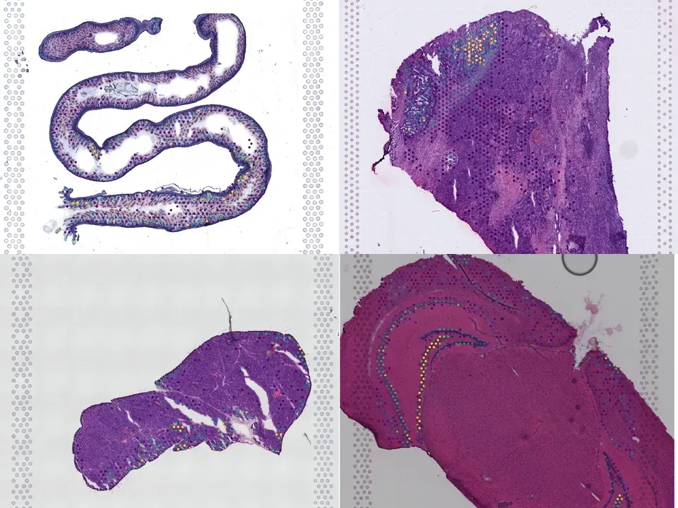
Mouse intestine
MEND32
mouse intestine 579 MB 1,970 spots silver H&E
TechnologyVisium
Organismmouse
Tissueintestine
Spots1,970
Magnification20x · 0.360 µm/px
silver H&E
from sddb import Catalog

cohort = Catalog().collection("doi-10-1038-s44161-022-00138-1")
cohort.members        # the 8 datasets as a table

print(cohort.licenses())              # check the terms before you download

sdatas = cohort.to_dict()             # {uid: SpatialData}, cached locally
# or keep the .zarr files in a folder of your choice:
# sdatas = cohort.to_dict(dest="data/")Needs pip install lamindb and network access to the Lamin database, which is limited during the alpha. If it fails, use the Client tab.
import lamindb as ln

db = ln.DB("scverse/spatialdata-db")
coll = db.Collection.get(key="doi-10.1038/s44161-022-00138-1")
coll.ordered_artifacts.to_dataframe()   # the 8 members, ordered

# download every member .zarr to the local cache:
paths = [a.cache() for a in coll.ordered_artifacts.all()]# sddb client: download every member .zarr into data/
sddb-cli download --collection doi-10-1038-s44161-022-00138-1 --dest data/Licenses: 8 CC BY-NC-SA 4.0
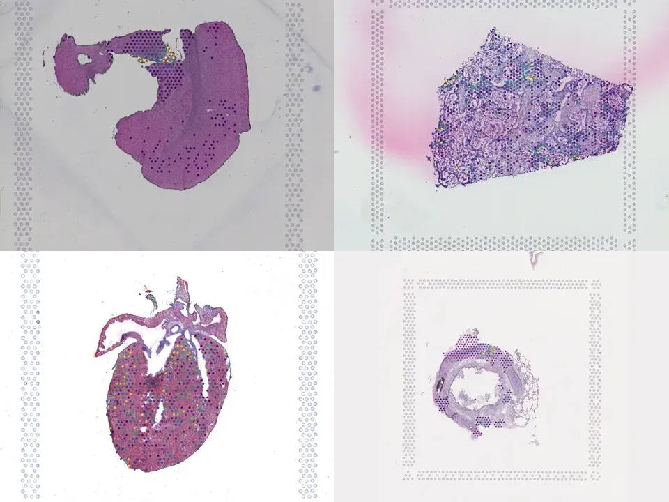
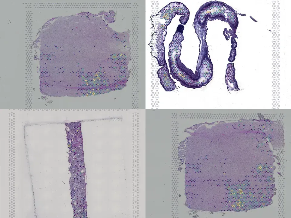
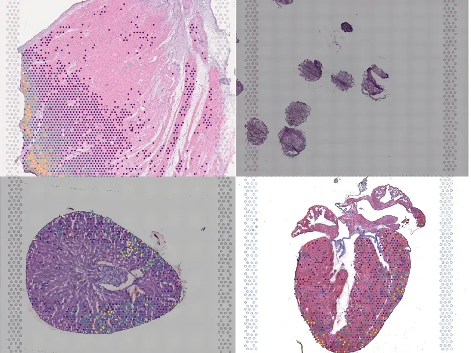
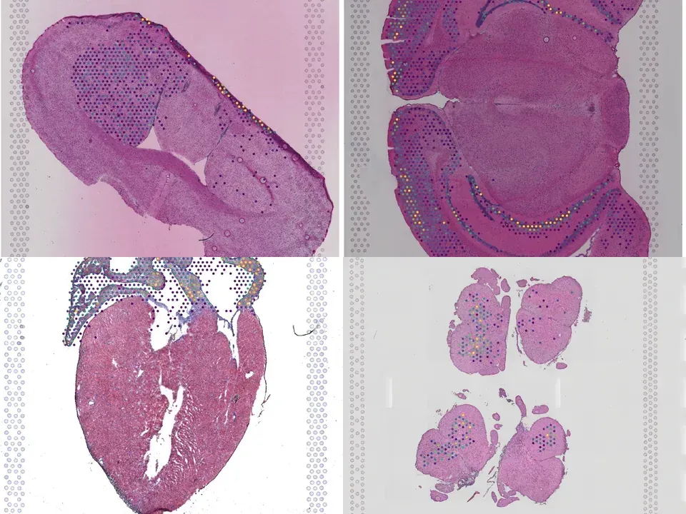
A significant fraction of sudden death in children and young adults is due to viral myocarditis, an inflammatory disease of the heart. In this study, by using integrated single-cell and spatial transcriptomics, we created a high-resolution, spatially resolved transcriptome map of reovirus-induced myocarditis in neonatal mouse hearts. We assayed hearts collected at three timepoints after infection and studied the temporal, spatial and cellular heterogeneity of host-virus interactions. We further assayed the intestine, the primary site of reovirus infection, to establish a full chronology of molecular events that ultimately lead to myocarditis. We found that inflamed endothelial cells recruit cytotoxic T cells and undergo pyroptosis in the myocarditic tissue. Analyses of spatially restricted gene expression in myocarditic regions and the border zone identified immune-mediated cell-type-specific injury and stress responses. Overall, we observed a complex network of cellular phenotypes and spatially restricted cell-cell interactions associated with reovirus-induced myocarditis in neonatal mice.
@article{Mantri_2022,
  title = {Spatiotemporal transcriptomics reveals pathogenesis of viral myocarditis},
  volume = {1},
  ISSN = {2731-0590},
  url = {http://dx.doi.org/10.1038/s44161-022-00138-1},
  DOI = {10.1038/s44161-022-00138-1},
  number = {10},
  journal = {Nature Cardiovascular Research},
  publisher = {Springer Science and Business Media LLC},
  author = {Mantri, Madhav and Hinchman, Meleana M. and McKellar, David W. and Wang, Michael F. Z. and Cross, Shaun T. and Parker, John S. L. and De Vlaminck, Iwijn},
  year = {2022},
  month = Oct,
  pages = {946–960}
}