
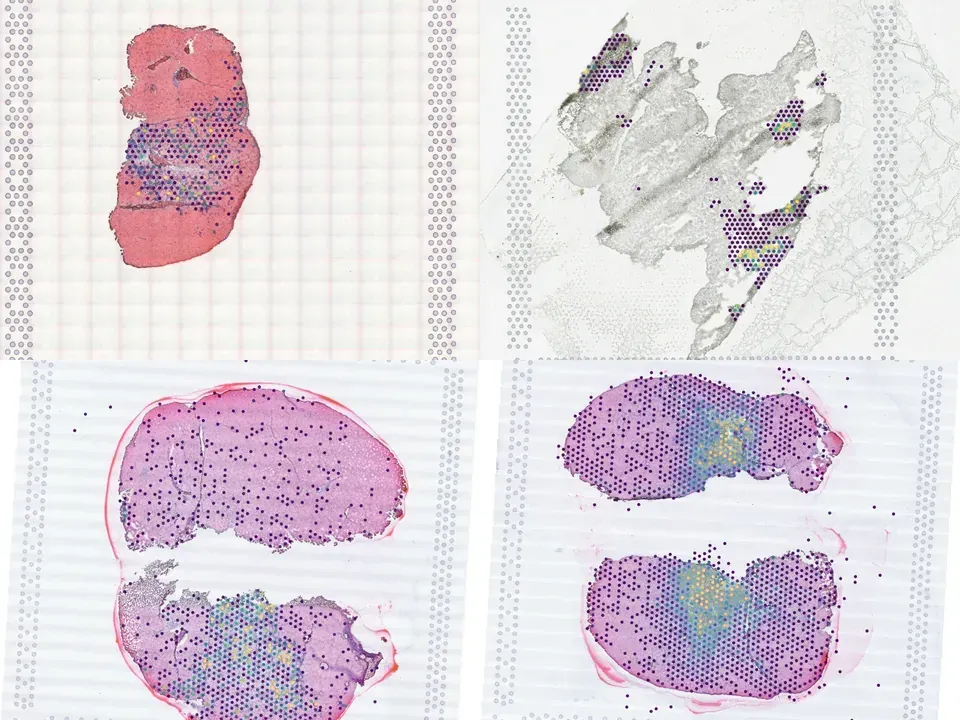
Mouse skeletal muscle tissue
NCBI796
mouse skeletal muscle tissue 1.1 GB 982 spots bronze H&E
TechnologyVisium
Organismmouse
Tissueskeletal muscle tissue
Spots982
Magnification20x · 0.360 µm/px
bronze H&E
from sddb import Catalog

cohort = Catalog().collection("doi-10-1038-s42003-021-02810-x")
cohort.members        # the 3 datasets as a table

print(cohort.licenses())              # check the terms before you download

sdatas = cohort.to_dict()             # {uid: SpatialData}, cached locally
# or keep the .zarr files in a folder of your choice:
# sdatas = cohort.to_dict(dest="data/")Needs pip install lamindb and network access to the Lamin database, which is limited during the alpha. If it fails, use the Client tab.
import lamindb as ln

db = ln.DB("scverse/spatialdata-db")
coll = db.Collection.get(key="doi-10.1038/s42003-021-02810-x")
coll.ordered_artifacts.to_dataframe()   # the 3 members, ordered

# download every member .zarr to the local cache:
paths = [a.cache() for a in coll.ordered_artifacts.all()]# sddb client: download every member .zarr into data/
sddb-cli download --collection doi-10-1038-s42003-021-02810-x --dest data/Licenses: 3 CC BY-NC-SA 4.0
Skeletal muscle repair is driven by the coordinated self-renewal and fusion of myogenic stem and progenitor cells. Single-cell gene expression analyses of myogenesis have been hampered by the poor sampling of rare and transient cell states that are critical for muscle repair, and do not inform the spatial context that is important for myogenic differentiation. Here, we demonstrate how large-scale integration of single-cell and spatial transcriptomic data can overcome these limitations. We created a single-cell transcriptomic dataset of mouse skeletal muscle by integration, consensus annotation, and analysis of 23 newly collected scRNAseq datasets and 88 publicly available single-cell (scRNAseq) and single-nucleus (snRNAseq) RNA-sequencing datasets. The resulting dataset includes more than 365,000 cells and spans a wide range of ages, injury, and repair conditions. Together, these data enabled identification of the predominant cell types in skeletal muscle, and resolved cell subtypes, including endothelial subtypes distinguished by vessel-type of origin, fibro-adipogenic progenitors defined by functional roles, and many distinct immune populations. The representation of different experimental conditions and the depth of transcriptome coverage enabled robust profiling of sparsely expressed genes. We built a densely sampled transcriptomic model of myogenesis, from stem cell quiescence to myofiber maturation, and identified rare, transitional states of progenitor commitment and fusion that are poorly represented in individual datasets. We performed spatial RNA sequencing of mouse muscle at three time points after injury and used the integrated dataset as a reference to achieve a high-resolution, local deconvolution of cell subtypes. We also used the integrated dataset to explore ligand-receptor co-expression patterns and identify dynamic cell-cell interactions in muscle injury response. We provide a public web tool to enable interactive exploration and visualization of the data. Our work supports the utility of large-scale integration of single-cell transcriptomic data as a tool for biological discovery.
@article{McKellar_2021,
  title = {Large-scale integration of single-cell transcriptomic data captures transitional progenitor states in mouse skeletal muscle regeneration},
  volume = {4},
  ISSN = {2399-3642},
  url = {http://dx.doi.org/10.1038/s42003-021-02810-x},
  DOI = {10.1038/s42003-021-02810-x},
  number = {1},
  journal = {Communications Biology},
  publisher = {Springer Science and Business Media LLC},
  author = {McKellar, David W. and Walter, Lauren D. and Song, Leo T. and Mantri, Madhav and Wang, Michael F. Z. and De Vlaminck, Iwijn and Cosgrove, Benjamin D.},
  year = {2021},
  month = Nov
}