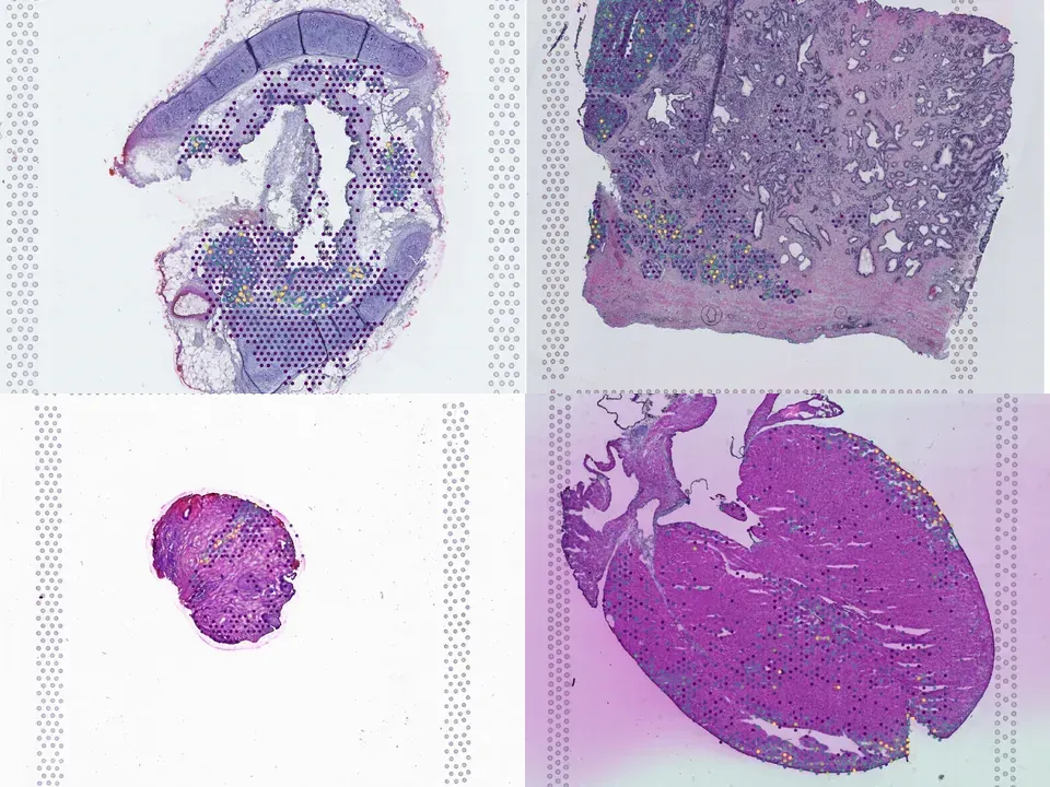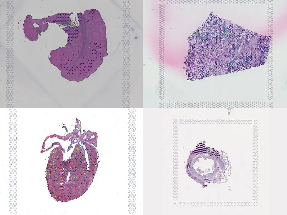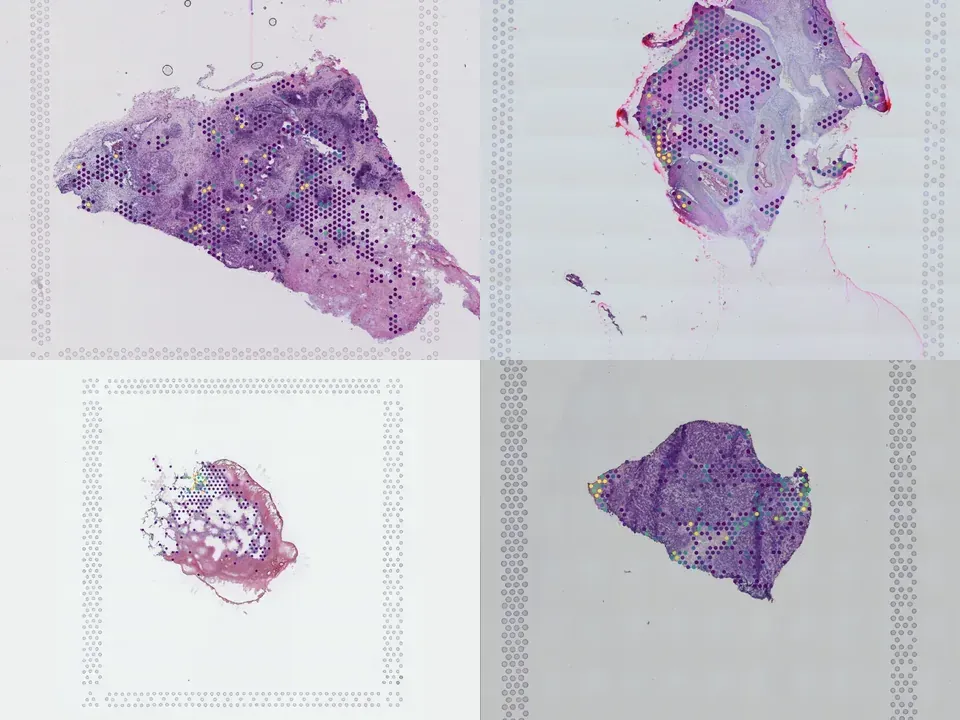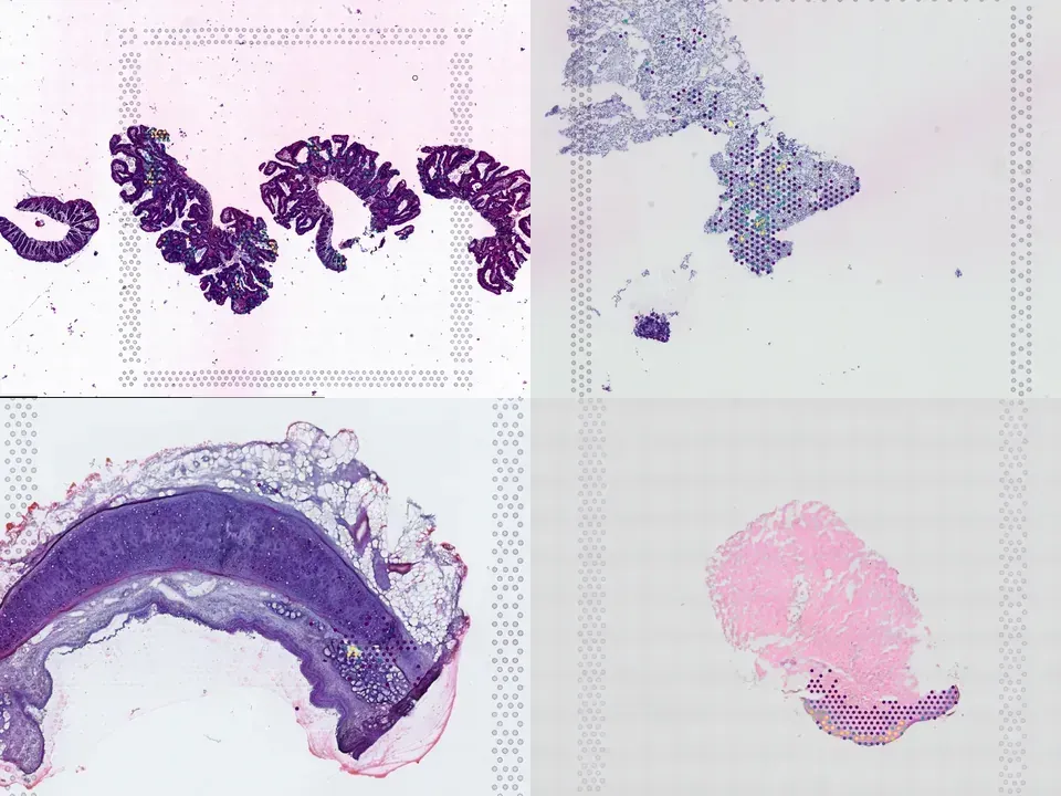
Collections / A spatially resolved atlas of the human lung characterizes a gland-associated immune niche

A spatially resolved atlas of the human lung characterizes a gland-associated immune niche

Datasets20
Size~9.6 GB
AssayVisium
Samplehuman · lung
KindPublication
Flag a data issue
10.1038/s41588-022-01243-4 ↗
Python · sddb client
from sddb import Catalog

cohort = Catalog().collection("doi-10-1038-s41588-022-01243-4")
cohort.members        # the 20 datasets as a table

print(cohort.licenses())              # check the terms before you download

sdatas = cohort.to_dict()             # {uid: SpatialData}, cached locally
# or keep the .zarr files in a folder of your choice:
# sdatas = cohort.to_dict(dest="data/")
Python · lamindb

Needs pip install lamindb and network access to the Lamin database, which is limited during the alpha. If it fails, use the Client tab.

import lamindb as ln

db = ln.DB("scverse/spatialdata-db")
coll = db.Collection.get(key="doi-10.1038/s41588-022-01243-4")
coll.ordered_artifacts.to_dataframe()   # the 20 members, ordered

# download every member .zarr to the local cache:
paths = [a.cache() for a in coll.ordered_artifacts.all()]
Shell
# sddb client: download every member .zarr into data/
sddb-cli download --collection doi-10-1038-s41588-022-01243-4 --dest data/

Licenses: 20 CC BY-NC-SA 4.0

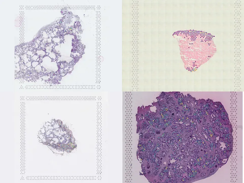
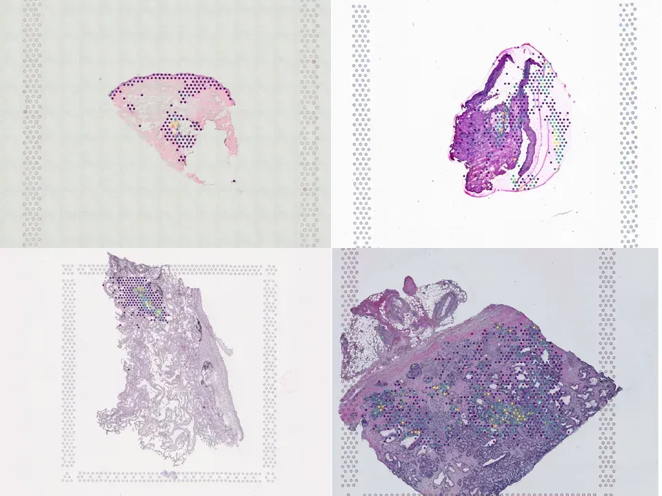
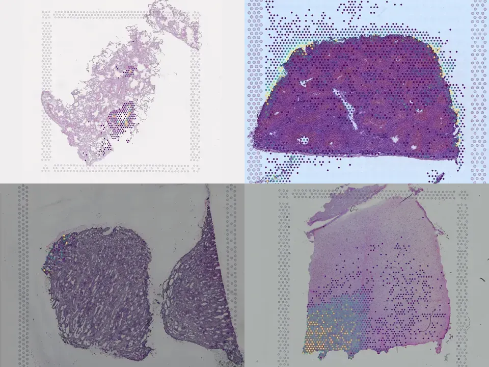
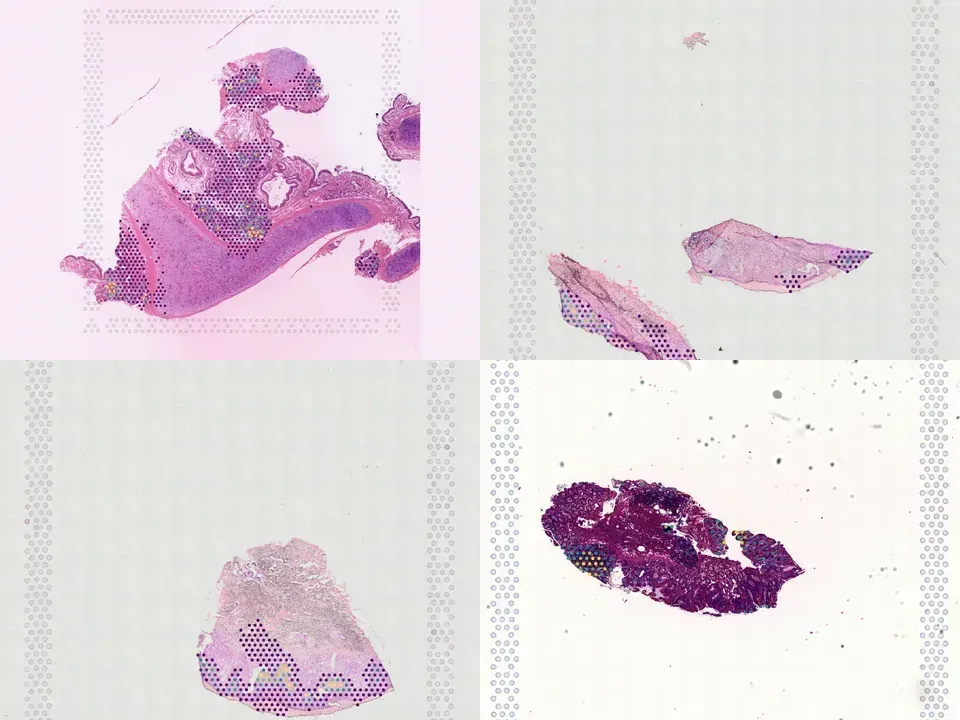
A spatially resolved atlas of the human lung characterizes a gland-associated immune niche.

Single-cell transcriptomics has allowed unprecedented resolution of cell types/states in the human lung, but their spatial context is less well defined. To (re)define tissue architecture of lung and airways, we profiled five proximal-to-distal locations of healthy human lungs in depth using multi-omic single cell/nuclei and spatial transcriptomics (queryable at lungcellatlas.org ). Using computational data integration and analysis, we extend beyond the suspension cell paradigm and discover macro and micro-anatomical tissue compartments including previously unannotated cell types in the epithelial, vascular, stromal and nerve bundle micro-environments. We identify and implicate peribronchial fibroblasts in lung disease. Importantly, we discover and validate a survival niche for IgA plasma cells in the airway submucosal glands (SMG). We show that gland epithelial cells recruit B cells and IgA plasma cells, and promote longevity and antibody secretion locally through expression of CCL28, APRIL and IL-6. This new 'gland-associated immune niche' has implications for respiratory health.
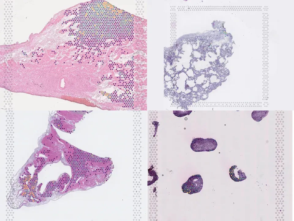
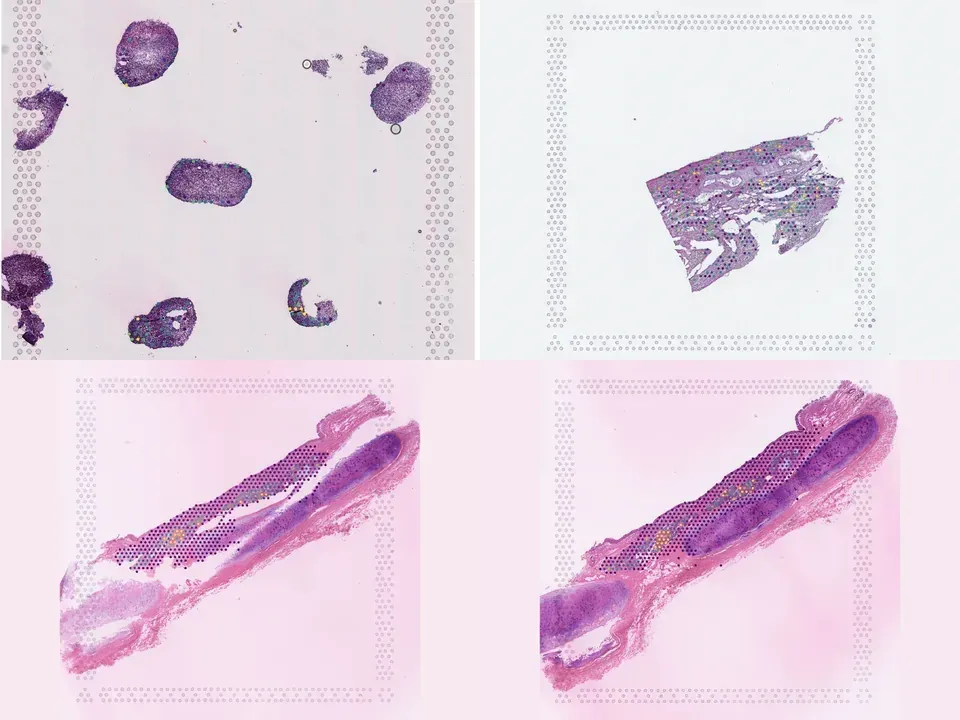
Open in Vitessce
Visium
Human lung
MISC15
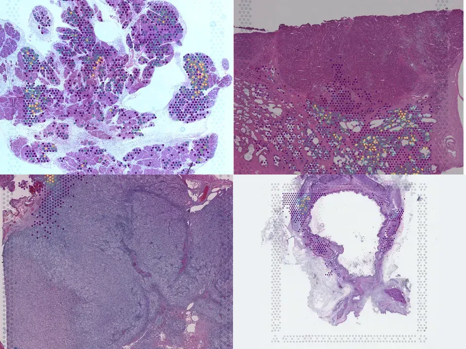
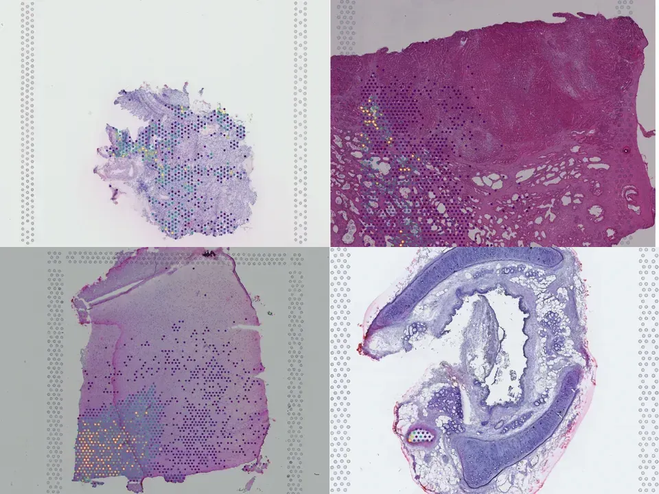
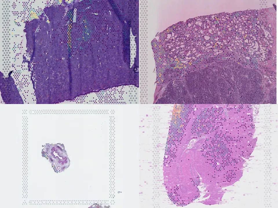
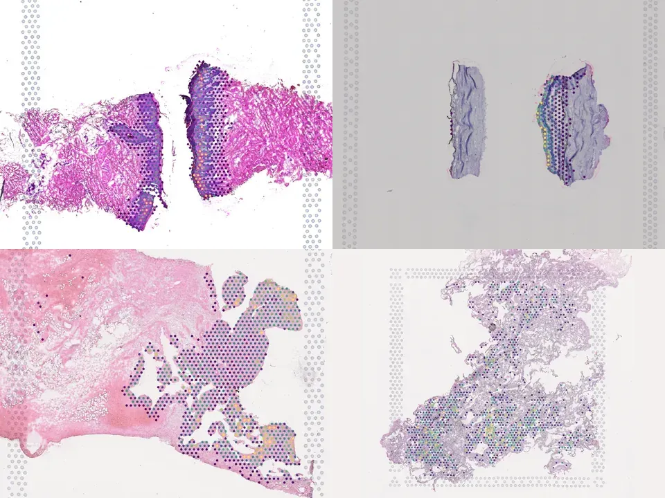
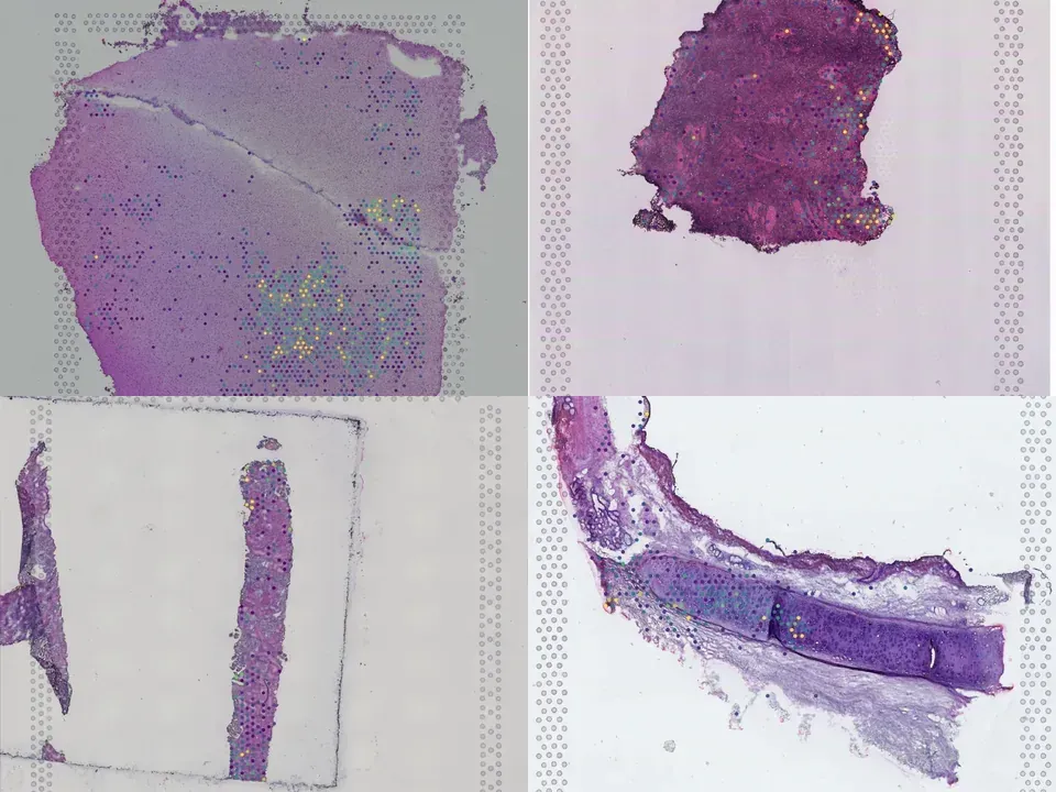
human lung 508 MB 2,501 spots silver H&E
TechnologyVisium
Organismhuman
Tissuelung
Size508 MB
Spots2,501
Magnification20x · 0.455 µm/px
silver H&E Open in Vitessce
Open in Vitessce
Visium
Human lung
MISC25
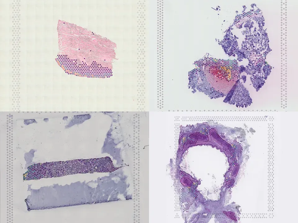
human lung 421 MB 2,249 spots silver H&E
TechnologyVisium
Organismhuman
Tissuelung
Size421 MB
Spots2,249
Magnification20x · 0.456 µm/px
silver H&E Open in Vitessce
Open in Vitessce
Visium
Human lung
MISC14
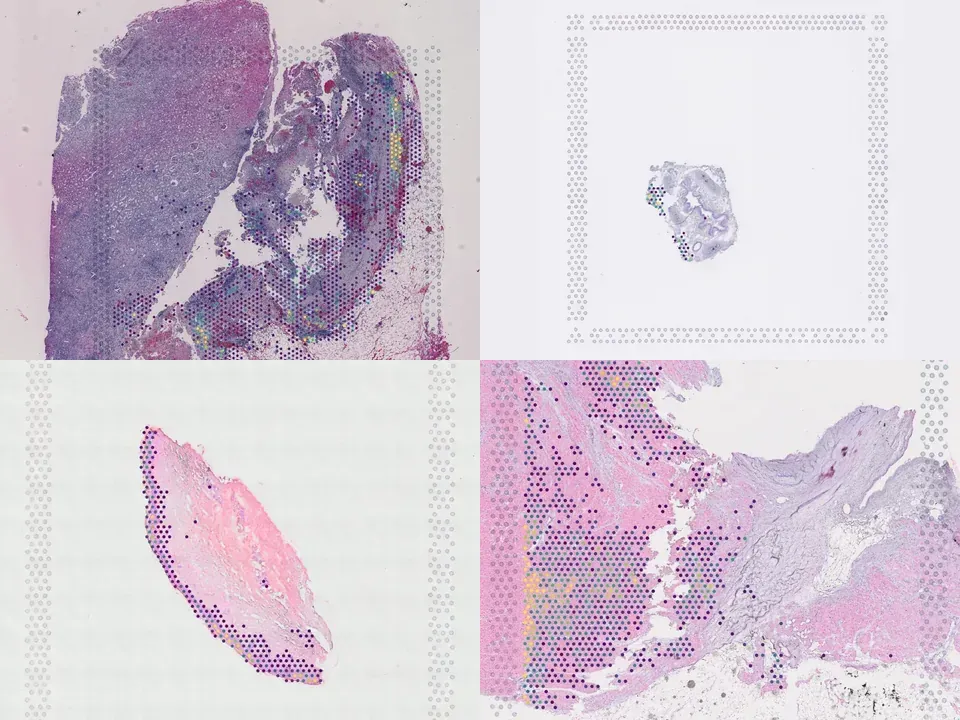
human lung 328 MB 1,670 spots silver H&E
TechnologyVisium
Organismhuman
Tissuelung
Size328 MB
Spots1,670
Magnification20x · 0.455 µm/px
silver H&E Open in Vitessce
Open in Vitessce
Visium
Human lung
MISC22
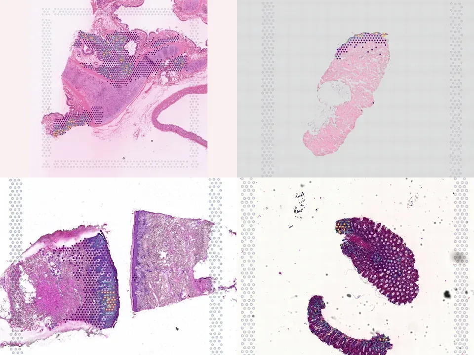
human lung 537 MB 2,704 spots silver H&E
TechnologyVisium
Organismhuman
Tissuelung
Size537 MB
Spots2,704
Magnification20x · 0.457 µm/px
silver H&E Open in Vitessce
Open in Vitessce
Visium
Human lung
MISC26
human lung 552 MB 2,524 spots silver H&E
TechnologyVisium
Organismhuman
Tissuelung
Size552 MB
Spots2,524
Magnification20x · 0.456 µm/px
silver H&E Open in Vitessce
Open in Vitessce
Visium
Human lung
MISC27
human lung 129 MB 408 spots silver H&E
TechnologyVisium
Organismhuman
Tissuelung
Size129 MB
Spots408
Magnification20x · 0.456 µm/px
silver H&E Open in Vitessce
Open in Vitessce
Visium
Human lung
MISC16
human lung 587 MB 3,154 spots silver H&E
TechnologyVisium
Organismhuman
Tissuelung
Size587 MB
Spots3,154
Magnification20x · 0.455 µm/px
silver H&E Open in Vitessce
Open in Vitessce
Visium
Human lung
MISC30
human lung 481 MB 2,729 spots silver H&E
TechnologyVisium
Organismhuman
Tissuelung
Size481 MB
Spots2,729
Magnification20x · 0.456 µm/px
silver H&E Open in Vitessce
Open in Vitessce
Visium
Human lung
MISC23
human lung 156 MB 436 spots silver H&E
TechnologyVisium
Organismhuman
Tissuelung
Size156 MB
Spots436
Magnification20x · 0.456 µm/px
silver H&E Open in Vitessce
Open in Vitessce
Visium
Human lung
MISC13
human lung 363 MB 1,768 spots silver H&E
TechnologyVisium
Organismhuman
Tissuelung
Size363 MB
Spots1,768
Magnification20x · 0.455 µm/px
silver H&E Open in Vitessce
Open in Vitessce
Visium
Human lung
MISC20
human lung 973 MB 3,316 spots silver H&E
TechnologyVisium
Organismhuman
Tissuelung
Size973 MB
Spots3,316
Magnification20x · 0.438 µm/px
silver H&E Open in Vitessce
Open in Vitessce
Visium
Human lung
MISC32
human lung 388 MB 1,928 spots silver H&E
TechnologyVisium
Organismhuman
Tissuelung
Size388 MB
Spots1,928
Magnification20x · 0.456 µm/px
silver H&E Open in Vitessce
Open in Vitessce
Visium
Human lung
MISC31
human lung 408 MB 2,524 spots silver H&E
TechnologyVisium
Organismhuman
Tissuelung
Size408 MB
Spots2,524
Magnification20x · 0.456 µm/px
silver H&E Open in Vitessce
Open in Vitessce
Visium
Human lung
MISC17
human lung 791 MB 1,084 spots silver H&E
TechnologyVisium
Organismhuman
Tissuelung
Size791 MB
Spots1,084
Magnification20x · 0.441 µm/px
silver H&E Open in Vitessce
Open in Vitessce
Visium
Human lung
MISC24
human lung 208 MB 875 spots silver H&E
TechnologyVisium
Organismhuman
Tissuelung
Size208 MB
Spots875
Magnification20x · 0.456 µm/px
silver H&E Open in Vitessce
Open in Vitessce
Visium
Human lung
MISC29
human lung 409 MB 2,283 spots silver H&E
TechnologyVisium
Organismhuman
Tissuelung
Size409 MB
Spots2,283
Magnification20x · 0.456 µm/px
silver H&E Open in Vitessce
Open in Vitessce
Visium
Human lung
MISC21
human lung 477 MB 2,987 spots silver H&E
TechnologyVisium
Organismhuman
Tissuelung
Size477 MB
Spots2,987
Magnification20x · 0.456 µm/px
silver H&E Open in Vitessce
Open in Vitessce
Visium
Human lung
MISC28
human lung 210 MB 863 spots silver H&E
TechnologyVisium
Organismhuman
Tissuelung
Size210 MB
Spots863
Magnification20x · 0.456 µm/px
silver H&E Open in Vitessce
Open in Vitessce
Visium
Human lung
MISC19
human lung 846 MB 2,454 spots silver H&E
TechnologyVisium
Organismhuman
Tissuelung
Size846 MB
Spots2,454
Magnification20x · 0.441 µm/px
silver H&E Open in Vitessce
Open in Vitessce
Visium
Human lung
MISC18
human lung 818 MB 2,081 spots silver H&E
TechnologyVisium
Organismhuman
Tissuelung
Size818 MB
Spots2,081
Magnification20x · 0.442 µm/px
silver H&E Open in Vitessce

Cite this collection

doi.org/10.1038/s41588-022-01243-4 ↗
BibTeX
@article{Madissoon_2022,
  title = {A spatially resolved atlas of the human lung characterizes a gland-associated immune niche},
  volume = {55},
  ISSN = {1546-1718},
  url = {http://dx.doi.org/10.1038/s41588-022-01243-4},
  DOI = {10.1038/s41588-022-01243-4},
  number = {1},
  journal = {Nature Genetics},
  publisher = {Springer Science and Business Media LLC},
  author = {Madissoon, Elo and Oliver, Amanda J. and Kleshchevnikov, Vitalii and Wilbrey-Clark, Anna and Polanski, Krzysztof and Richoz, Nathan and Ribeiro Orsi, Ana and Mamanova, Lira and Bolt, Liam and Elmentaite, Rasa and Pett, J. Patrick and Huang, Ni and Xu, Chuan and He, Peng and Dabrowska, Monika and Pritchard, Sophie and Tuck, Liz and Prigmore, Elena and Perera, Shani and Knights, Andrew and Oszlanczi, Agnes and Hunter, Adam and Vieira, Sara F. and Patel, Minal and Lindeboom, Rik G. H. and Campos, Lia S. and Matsuo, Kazuhiko and Nakayama, Takashi and Yoshida, Masahiro and Worlock, Kaylee B. and Nikolić, Marko Z. and Georgakopoulos, Nikitas and Mahbubani, Krishnaa T. and Saeb-Parsy, Kourosh and Bayraktar, Omer Ali and Clatworthy, Menna R. and Stegle, Oliver and Kumasaka, Natsuhiko and Teichmann, Sarah A. and Meyer, Kerstin B.},
  year = {2022},
  month = Dec,
  pages = {66–77}
}