
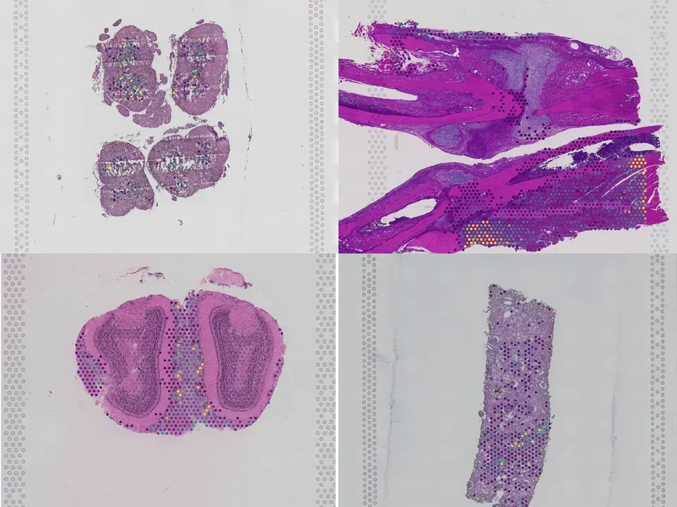
Mouse spinal cord
NCBI445
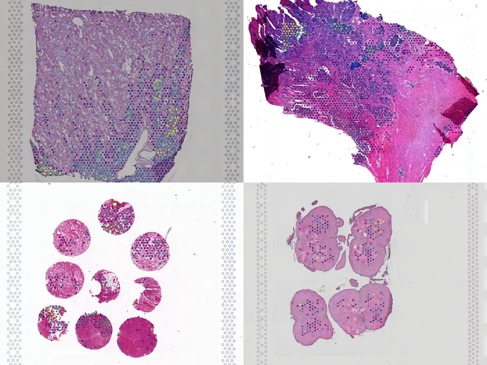
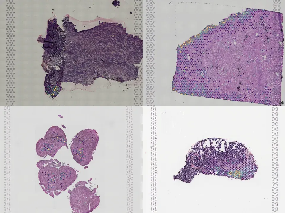
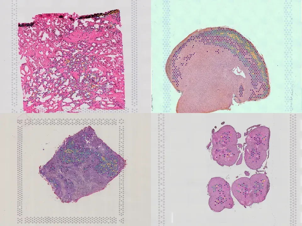
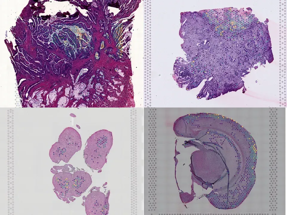
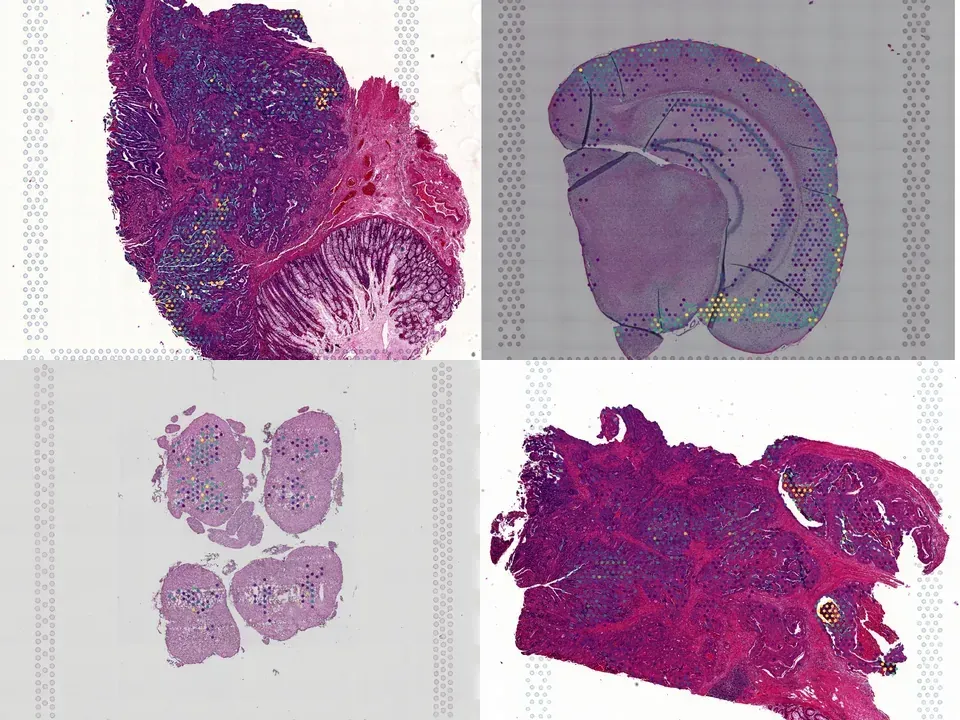
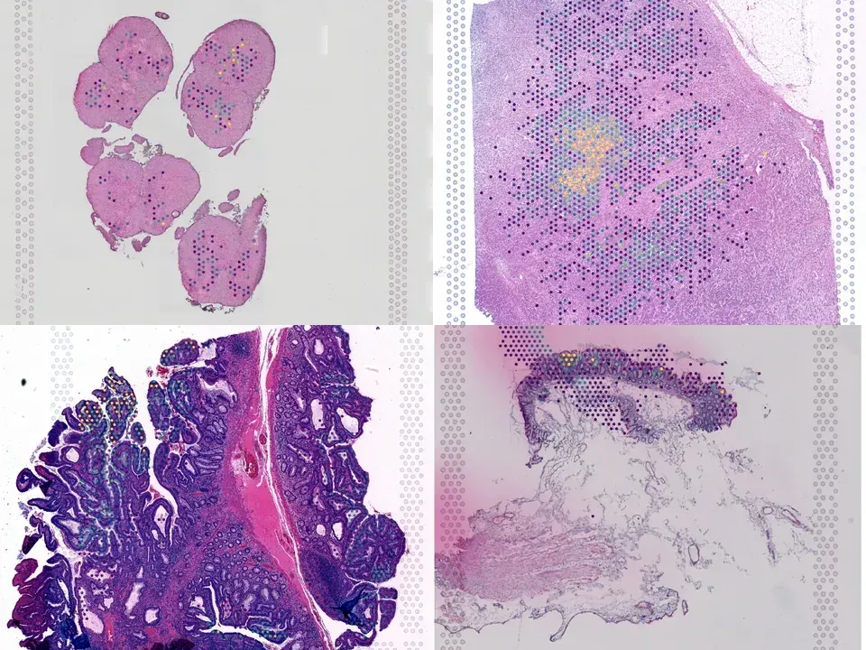
mouse spinal cord 296 MB 556 spots silver H&E
TechnologyVisium
Organismmouse
Tissuespinal cord
Spots556
silver H&E
from sddb import Catalog

cohort = Catalog().collection("doi-10-1038-s41586-022-05385-7")
cohort.members        # the 16 datasets as a table

print(cohort.licenses())              # check the terms before you download

sdatas = cohort.to_dict()             # {uid: SpatialData}, cached locally
# or keep the .zarr files in a folder of your choice:
# sdatas = cohort.to_dict(dest="data/")Needs pip install lamindb and network access to the Lamin database, which is limited during the alpha. If it fails, use the Client tab.
import lamindb as ln

db = ln.DB("scverse/spatialdata-db")
coll = db.Collection.get(key="doi-10.1038/s41586-022-05385-7")
coll.ordered_artifacts.to_dataframe()   # the 16 members, ordered

# download every member .zarr to the local cache:
paths = [a.cache() for a in coll.ordered_artifacts.all()]# sddb client: download every member .zarr into data/
sddb-cli download --collection doi-10-1038-s41586-022-05385-7 --dest data/Licenses: 16 CC BY-NC-SA 4.0
A spinal cord injury interrupts pathways from the brain and brainstem that project to the lumbar spinal cord, leading to paralysis. Here we show that spatiotemporal epidural electrical stimulation (EES) of the lumbar spinal cord<sup>1-3</sup> applied during neurorehabilitation<sup>4,5</sup> (EES<sup>REHAB</sup>) restored walking in nine individuals with chronic spinal cord injury. This recovery involved a reduction in neuronal activity in the lumbar spinal cord of humans during walking. We hypothesized that this unexpected reduction reflects activity-dependent selection of specific neuronal subpopulations that become essential for a patient to walk after spinal cord injury. To identify these putative neurons, we modelled the technological and therapeutic features underlying EES<sup>REHAB</sup> in mice. We applied single-nucleus RNA sequencing<sup>6-9</sup> and spatial transcriptomics<sup>10,11</sup> to the spinal cords of these mice to chart a spatially resolved molecular atlas of recovery from paralysis. We then employed cell type<sup>12,13</sup> and spatial prioritization to identify the neurons involved in the recovery of walking. A single population of excitatory interneurons nested within intermediate laminae emerged. Although these neurons are not required for walking before spinal cord injury, we demonstrate that they are essential for the recovery of walking with EES following spinal cord injury. Augmenting the activity of these neurons phenocopied the recovery of walking enabled by EES<sup>REHAB</sup>, whereas ablating them prevented the recovery of walking that occurs spontaneously after moderate spinal cord injury. We thus identified a recovery-organizing neuronal subpopulation that is necessary and sufficient to regain walking after paralysis. Moreover, our methodology establishes a framework for using molecular cartography to identify the neurons that produce complex behaviours.
@article{Kathe_2022,
  title = {The neurons that restore walking after paralysis},
  volume = {611},
  ISSN = {1476-4687},
  url = {http://dx.doi.org/10.1038/s41586-022-05385-7},
  DOI = {10.1038/s41586-022-05385-7},
  number = {7936},
  journal = {Nature},
  publisher = {Springer Science and Business Media LLC},
  author = {Kathe, Claudia and Skinnider, Michael A. and Hutson, Thomas H. and Regazzi, Nicola and Gautier, Matthieu and Demesmaeker, Robin and Komi, Salif and Ceto, Steven and James, Nicholas D. and Cho, Newton and Baud, Laetitia and Galan, Katia and Matson, Kaya J. E. and Rowald, Andreas and Kim, Kyungjin and Wang, Ruijia and Minassian, Karen and Prior, John O. and Asboth, Leonie and Barraud, Quentin and Lacour, Stéphanie P. and Levine, Ariel J. and Wagner, Fabien and Bloch, Jocelyne and Squair, Jordan W. and Courtine, Grégoire},
  year = {2022},
  month = Nov,
  pages = {540–547}
}