
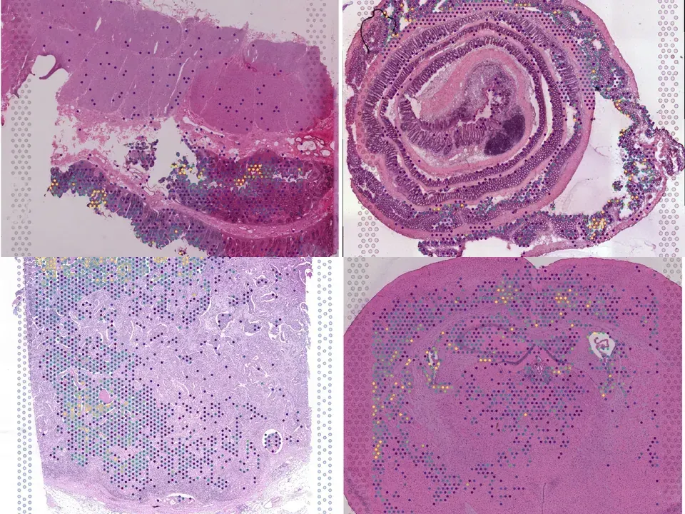
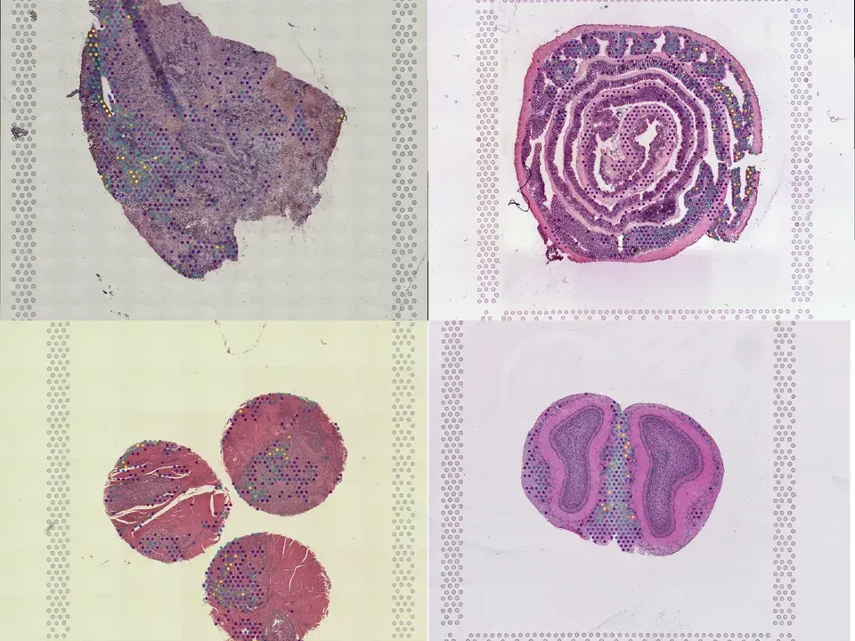
Mouse intestine
NCBI728
mouse intestine ulcerative colitis 269 MB 3,630 spots silver H&E
TechnologyVisium
Organismmouse
Tissueintestine
Conditionulcerative colitis
Spots3,630
silver H&E
from sddb import Catalog

cohort = Catalog().collection("doi-10-1016-j-crmeth-2022-100340")
cohort.members        # the 2 datasets as a table

print(cohort.licenses())              # check the terms before you download

sdatas = cohort.to_dict()             # {uid: SpatialData}, cached locally
# or keep the .zarr files in a folder of your choice:
# sdatas = cohort.to_dict(dest="data/")Needs pip install lamindb and network access to the Lamin database, which is limited during the alpha. If it fails, use the Client tab.
import lamindb as ln

db = ln.DB("scverse/spatialdata-db")
coll = db.Collection.get(key="doi-10.1016/j.crmeth.2022.100340")
coll.ordered_artifacts.to_dataframe()   # the 2 members, ordered

# download every member .zarr to the local cache:
paths = [a.cache() for a in coll.ordered_artifacts.all()]# sddb client: download every member .zarr into data/
sddb-cli download --collection doi-10-1016-j-crmeth-2022-100340 --dest data/Licenses: 2 CC BY-NC-SA 4.0
Tumor heterogeneity is a major challenge for oncology drug discovery and development. Understanding of the spatial tumor landscape is key to identifying new targets and impactful model systems. Here, we test the utility of spatial transcriptomics (ST) for oncology discovery by profiling 40 tissue sections and 80,024 capture spots across a diverse set of tissue types, sample formats, and RNA capture chemistries. We verify the accuracy and fidelity of ST by leveraging matched pathology analysis, which provides a ground truth for tissue section composition. We then use spatial data to demonstrate the capture of key tumor depth features, identifying hypoxia, necrosis, vasculature, and extracellular matrix variation. We also leverage spatial context to identify relative cell-type locations showing the anti-correlation of tumor and immune cells in syngeneic cancer models. Lastly, we demonstrate target identification approaches in clinical pancreatic adenocarcinoma samples, highlighting tumor intrinsic biomarkers and paracrine signaling.
@article{Lyubetskaya_2022,
  title = {Assessment of spatial transcriptomics for oncology discovery},
  volume = {2},
  ISSN = {2667-2375},
  url = {http://dx.doi.org/10.1016/j.crmeth.2022.100340},
  DOI = {10.1016/j.crmeth.2022.100340},
  number = {11},
  journal = {Cell Reports Methods},
  publisher = {Elsevier BV},
  author = {Lyubetskaya, Anna and Rabe, Brian and Fisher, Andrew and Lewin, Anne and Neuhaus, Isaac and Brett, Constance and Brett, Todd and Pereira, Ethel and Golhar, Ryan and Kebede, Sami and Font-Tello, Alba and Mosure, Kathy and Van Wittenberghe, Nicholas and Mavrakis, Konstantinos J. and MacIsaac, Kenzie and Chen, Benjamin J. and Drokhlyansky, Eugene},
  year = {2022},
  month = Nov,
  pages = {100340}
}